Identification of Novel GSK-3 beta Hits Using Competitive Biophysical Assays.
Balboni, B., Tripathi, S.K., Veronesi, M., Russo, D., Penna, I., Giabbai, B., Bandiera, T., Storici, P., Girotto, S., Cavalli, A.(2022) Int J Mol Sci 23
- PubMed: 35409221 Search on PubMedSearch on PubMed Central
- DOI: https://doi.org/10.3390/ijms23073856
- Primary Citation Related Structures:
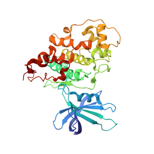
7U2Z, 7U31, 7U33, 7U36 - PubMed Abstract:
Glycogen synthase kinase 3 beta (GSK-3β) is an evolutionarily conserved serine-threonine kinase dysregulated in numerous pathologies, such as Alzheimer's disease and cancer. Even though GSK-3β is a validated pharmacological target most of its inhibitors have two main limitations: the lack of selectivity due to the high homology that characterizes the ATP binding site of most kinases, and the toxicity that emerges from GSK-3β complete inhibition which translates into the impairment of the plethora of pathways GSK-3β is involved in. Starting from a 1D 19 F NMR fragment screening, we set up several biophysical assays for the identification of GSK-3β inhibitors capable of binding protein hotspots other than the ATP binding pocket or to the ATP binding pocket, but with an affinity able of competing with a reference binder. A phosphorylation activity assay on a panel of several kinases provided selectivity data that were further rationalized and corroborated by structural information on GSK-3β in complex with the hit compounds. In this study, we identified promising fragments, inhibitors of GSK-3β, while proposing an alternative screening workflow that allows facing the flaws that characterize the most common GSK-3β inhibitors through the identification of selective inhibitors and/or inhibitors able to modulate GSK-3β activity without leading to its complete inhibition.
- Computational and Chemical Biology, Istituto Italiano di Tecnologia, Via Morego 30, 16163 Genova, Italy.
Organizational Affiliation: