7U0A | pdb_00007u0a

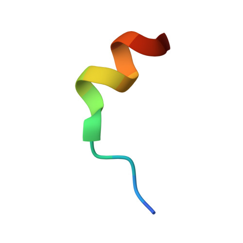
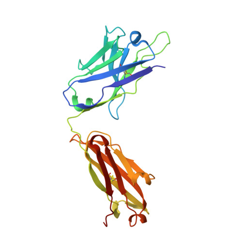
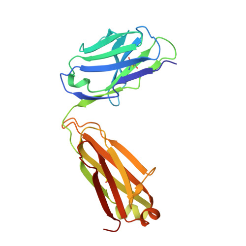
Crystal Structure of C77G12 Fab in complex with SARS-CoV-2 S fusion peptide


Experimental Data Snapshot

  • Method: X-RAY DIFFRACTION
  • Resolution: 1.70 Å
  • R-Value Free: 
    0.187 (Depositor), 0.187 (DCC) 
  • R-Value Work: 
    0.154 (Depositor), 0.154 (DCC) 
  • R-Value Observed: 
    0.156 (Depositor) 

Starting Model: experimental
View more details

wwPDB Validation 3D Report Full Report

Validation slider image for 7U0A

This is version 2.3 of the entry. See complete history

Literature

ACE2-binding exposes the SARS-CoV-2 fusion peptide to broadly neutralizing coronavirus antibodies.

Low, J.S.Jerak, J.Tortorici, M.A.McCallum, M.Pinto, D.Cassotta, A.Foglierini, M.Mele, F.Abdelnabi, R.Weynand, B.Noack, J.Montiel-Ruiz, M.Bianchi, S.Benigni, F.Sprugasci, N.Joshi, A.Bowen, J.E.Stewart, C.Rexhepaj, M.Walls, A.C.Jarrossay, D.Morone, D.Paparoditis, P.Garzoni, C.Ferrari, P.Ceschi, A.Neyts, J.Purcell, L.A.Snell, G.Corti, D.Lanzavecchia, A.Veesler, D.Sallusto, F.

(2022) Science 377: 735-742

  • DOI: https://doi.org/10.1126/science.abq2679
  • Primary Citation Related Structures: 
    7SKZ, 7SL5, 7U09, 7U0A, 7U0E

  • PubMed Abstract: 

    The coronavirus spike glycoprotein attaches to host receptors and mediates viral fusion. Using a broad screening approach, we isolated seven monoclonal antibodies (mAbs) that bind to all human-infecting coronavirus spike proteins from severe acute respiratory syndrome coronavirus 2 (SARS-CoV-2) immune donors. These mAbs recognize the fusion peptide and acquire affinity and breadth through somatic mutations. Despite targeting a conserved motif, only some mAbs show broad neutralizing activity in vitro against alpha- and betacoronaviruses, including animal coronaviruses WIV-1 and PDF-2180. Two selected mAbs also neutralize Omicron BA.1 and BA.2 authentic viruses and reduce viral burden and pathology in vivo. Structural and functional analyses showed that the fusion peptide-specific mAbs bound with different modalities to a cryptic epitope hidden in prefusion stabilized spike, which became exposed upon binding of angiotensin-converting enzyme 2 (ACE2) or ACE2-mimicking mAbs.


  • Organizational Affiliation
    • Institute for Research in Biomedicine, Università della Svizzera Italiana, 6500 Bellinzona, Switzerland.

Macromolecule Content 

  • Total Structure Weight: 48.98 kDa 
  • Atom Count: 4,071 
  • Modeled Residue Count: 451 
  • Deposited Residue Count: 453 
  • Unique protein chains: 3

Macromolecules

Find similar proteins by:  Sequence   |   3D Structure  
Entity ID: 1
MoleculeChains  Sequence LengthOrganismDetailsImage
SARS-CoV-2 S fusion peptide14Severe acute respiratory syndrome coronavirus 2Mutation(s): 0 
Find similar proteins by:|  3D Structure
Entity ID: 2
MoleculeChains  Sequence LengthOrganismDetailsImage
Heavy chain Fab C77G12B [auth H]225Homo sapiensMutation(s): 0 
Find similar proteins by:|  3D Structure
Entity ID: 3
MoleculeChains  Sequence LengthOrganismDetailsImage
Light chain Fab C77G12C [auth L]214Homo sapiensMutation(s): 0 

Small Molecules

Ligands 1 Unique
IDChains Name / Formula / InChI Key2D Diagram3D Interactions
CL

Query on CL


Download:Ideal Coordinates CCD File
D [auth H]CHLORIDE ION
Cl
VEXZGXHMUGYJMC-UHFFFAOYSA-M

Experimental Data & Validation

Experimental Data

  • Method: X-RAY DIFFRACTION
  • Resolution: 1.70 Å
  • R-Value Free:  0.187 (Depositor), 0.187 (DCC) 
  • R-Value Work:  0.154 (Depositor), 0.154 (DCC) 
  • R-Value Observed: 0.156 (Depositor) 
Space Group: P 1 21 1
Unit Cell:
Length ( Å )Angle ( ˚ )
a = 45.227α = 90
b = 64.056β = 93.33
c = 78.813γ = 90
Software Package:
Software NamePurpose
PHENIXrefinement
PDB_EXTRACTdata extraction
XDSdata reduction
Aimlessdata scaling
PHENIXphasing

Structure Validation

View Full Validation Report


Entry History 

& Funding Information

Deposition Data


Funding OrganizationLocationGrant Number
Partnership for Structural Biology (PSB)FranceSSGCID

Revision History  (Full details and data files)

  • Version 1.0: 2022-08-03
    Type: Initial release
  • Version 2.0: 2022-08-10
    Changes: Advisory, Atomic model, Database references, Derived calculations, Source and taxonomy, Structure summary
  • Version 2.1: 2022-08-24
    Changes: Database references
  • Version 2.2: 2023-10-18
    Changes: Data collection, Refinement description
  • Version 2.3: 2024-11-13
    Changes: Structure summary