7TUU | pdb_00007tuu

Structure of the SARS-CoV-2 main protease in complex with halicin


Experimental Data Snapshot

  • Method: X-RAY DIFFRACTION
  • Resolution: 1.85 Å
  • R-Value Free: 
    0.272 (Depositor), 0.272 (DCC) 
  • R-Value Work: 
    0.249 (Depositor), 0.250 (DCC) 
  • R-Value Observed: 
    0.251 (Depositor) 

Starting Model: experimental
View more details

wwPDB Validation 3D Report Full Report

Validation slider image for 7TUU

Ligand Structure Quality Assessment 


This is version 1.2 of the entry. See complete history

Literature

Repurposing Halicin as a potent covalent inhibitor for the SARS-CoV-2 main protease.

Yang, K.S.Alex Kuo, S.T.Blankenship, L.R.Geng, Z.Z.Li, S.G.Russell, D.H.Yan, X.Xu, S.Liu, W.R.

(2022) Curr Res Chem Biol 2: 100025-100025

  • DOI: https://doi.org/10.1016/j.crchbi.2022.100025
  • Primary Citation Related Structures: 
    7TUU

  • PubMed Abstract: 

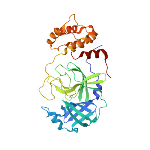
    The rapid spread of COVID-19 has caused a worldwide public health crisis. For prompt and effective development of antivirals for SARS-CoV-2, the pathogen of COVID-19, drug repurposing has been broadly conducted by targeting the main protease (M Pro ), a key enzyme responsible for the replication of virus inside the host. In this study, we evaluate the inhibition potency of a nitrothiazole-containing drug, halicin, and reveal its reaction and interaction mechanism with M Pro . The in vitro potency test shows that halicin inhibits the activity of M Pro an IC 50 of 181.7 ​nM. Native mass spectrometry and X-ray crystallography studies clearly indicate that the nitrothiazole fragment of halicin covalently binds to the catalytic cysteine C145 of M Pro . Interaction and conformational changes inside the active site of M Pro suggest a favorable nucleophilic aromatic substitution reaction mechanism between M Pro C145 and halicin, explaining the high inhibition potency of halicin towards M Pro .


  • Organizational Affiliation
    • Texas A&M Drug Discovery Laboratory, Department of Chemistry, Texas A&M University, College Station, TX, 77843, USA.

Macromolecule Content 

  • Total Structure Weight: 34.22 kDa 
  • Atom Count: 2,580 
  • Modeled Residue Count: 306 
  • Deposited Residue Count: 306 
  • Unique protein chains: 1

Macromolecules

Find similar proteins by:|  3D Structure
Entity ID: 1
MoleculeChains  Sequence LengthOrganismDetailsImage
3C-like proteinase nsp5306Severe acute respiratory syndrome coronavirus 2Mutation(s): 0 
EC: 3.4.22.69
UniProt
Find proteins for P0DTD1 (Severe acute respiratory syndrome coronavirus 2)
Explore P0DTD1 
Go to UniProtKB:  P0DTD1
Entity Groups
Sequence Clusters30% Identity50% Identity70% Identity90% Identity95% Identity100% Identity
UniProt GroupP0DTD1
Sequence Annotations
Expand
Reference Sequence

Experimental Data & Validation

Experimental Data

  • Method: X-RAY DIFFRACTION
  • Resolution: 1.85 Å
  • R-Value Free:  0.272 (Depositor), 0.272 (DCC) 
  • R-Value Work:  0.249 (Depositor), 0.250 (DCC) 
  • R-Value Observed: 0.251 (Depositor) 
Space Group: I 1 2 1
Unit Cell:
Length ( Å )Angle ( ˚ )
a = 52.213α = 90
b = 84.362β = 95.608
c = 90.093γ = 90
Software Package:
Software NamePurpose
PHENIXrefinement
PROTEUM PLUSdata reduction
Aimlessdata scaling
PHENIXphasing

Structure Validation

View Full Validation Report


Ligand Structure Quality Assessment 


Entry History 

& Funding Information

Deposition Data


Funding OrganizationLocationGrant Number
Welch FoundationUnited States--

Revision History  (Full details and data files)

  • Version 1.0: 2022-08-03
    Type: Initial release
  • Version 1.1: 2023-10-18
    Changes: Data collection, Refinement description
  • Version 1.2: 2024-11-20
    Changes: Structure summary