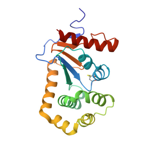
7TTV

E.coli DsbA in complex with 4-phenyl-2-(3-phenylpropyl)thiazole-5-carboxylic acid


Experimental Data Snapshot

  • Method: X-RAY DIFFRACTION
  • Resolution: 1.99 Å
  • R-Value Free: 0.227 
  • R-Value Work: 0.181 
  • R-Value Observed: 0.183 

wwPDB Validation   3D Report Full Report


Ligand Structure Quality Assessment 


This is version 1.1 of the entry. See complete history


Literature

Methyl probes in proteins for determining ligand binding mode in weak protein-ligand complexes.

Mohanty, B.Orts, J.Wang, G.Nebl, S.Alwan, W.S.Doak, B.C.Williams, M.L.Heras, B.Mobli, M.Scanlon, M.J.

(2022) Sci Rep 12: 11231-11231

  • DOI: https://doi.org/10.1038/s41598-022-13561-y
  • Primary Citation of Related Structures:  
    7TTV

  • PubMed Abstract: 

    Structures of protein-ligand complexes provide critical information for drug design. Most protein-ligand complex structures are determined using X-ray crystallography, but where crystallography is not able to generate a structure for a complex, NMR is often the best alternative. However, the available tools to enable rapid and robust structure determination of protein-ligand complexes by NMR are currently limited. This leads to situations where projects are either discontinued or pursued without structural data, rendering the task more difficult. We previously reported the NMR Molecular Replacement (NMR 2 ) approach that allows the structure of a protein-ligand complex to be determined without requiring the cumbersome task of protein resonance assignment. Herein, we describe the NMR 2  approach to determine the binding pose of a small molecule in a weak protein-ligand complex by collecting sparse protein methyl-to-ligand NOEs from a selectively labeled protein sample and an unlabeled ligand. In the selective labeling scheme all methyl containing residues of the protein are protonated in an otherwise deuterated background. This allows measurement of intermolecular NOEs with greater sensitivity using standard NOESY pulse sequences instead of isotope-filtered NMR experiments. This labelling approach is well suited to the NMR 2 approach and extends its utility to include larger protein-ligand complexes.


  • Organizational Affiliation

    Medicinal Chemistry, Monash Institute of Pharmaceutical Sciences, Monash University, 381 Royal Parade, Parkville, VIC, 3052, Australia.


Macromolecules
Find similar proteins by:  (by identity cutoff)  |  3D Structure
Entity ID: 1
MoleculeChains Sequence LengthOrganismDetailsImage
Thiol:disulfide interchange protein DsbA
A, B
189Escherichia coli K-12Mutation(s): 0 
Gene Names: dsbAdsfppfAb3860JW3832
UniProt
Find proteins for P0AEG4 (Escherichia coli (strain K12))
Explore P0AEG4 
Go to UniProtKB:  P0AEG4
Entity Groups  
Sequence Clusters30% Identity50% Identity70% Identity90% Identity95% Identity100% Identity
UniProt GroupP0AEG4
Sequence Annotations
Expand
  • Reference Sequence
Small Molecules
Ligands 2 Unique
IDChains Name / Formula / InChI Key2D Diagram3D Interactions
QVP (Subject of Investigation/LOI)
Query on QVP

Download Ideal Coordinates CCD File 
C [auth A]4-phenyl-2-(3-phenylpropyl)-1,3-thiazole-5-carboxylic acid
C19 H17 N O2 S
WWFYLUZIXCVYBZ-UHFFFAOYSA-N
CU
Query on CU

Download Ideal Coordinates CCD File 
D [auth B]COPPER (II) ION
Cu
JPVYNHNXODAKFH-UHFFFAOYSA-N
Experimental Data & Validation

Experimental Data

  • Method: X-RAY DIFFRACTION
  • Resolution: 1.99 Å
  • R-Value Free: 0.227 
  • R-Value Work: 0.181 
  • R-Value Observed: 0.183 
  • Space Group: C 1 2 1
Unit Cell:
Length ( Å )Angle ( ˚ )
a = 117.45α = 90
b = 63.74β = 125.87
c = 74.28γ = 90
Software Package:
Software NamePurpose
PHENIXrefinement
SCALAdata scaling
PDB_EXTRACTdata extraction
MOSFLMdata reduction
PHASERphasing

Structure Validation

View Full Validation Report


Ligand Structure Quality Assessment 


Entry History & Funding Information

Deposition Data


Funding OrganizationLocationGrant Number
National Health and Medical Research Council (NHMRC, Australia)AustraliaGNT1099151

Revision History  (Full details and data files)

  • Version 1.0: 2023-04-12
    Type: Initial release
  • Version 1.1: 2023-10-25
    Changes: Data collection, Refinement description