7TOL

X-ray crystal structure of glycerol dibiphytanyl glycerol tetraether - macrocyclic archaeol synthase (GDGT-MAS) from Methanocaldococcus jannaschii with archaeal lipid, 5'deoxyadenosine, and methionine bound


Experimental Data Snapshot

  • Method: X-RAY DIFFRACTION
  • Resolution: 2.03 Å
  • R-Value Free: 0.229 
  • R-Value Work: 0.186 
  • R-Value Observed: 0.189 

wwPDB Validation   3D Report Full Report


Ligand Structure Quality Assessment 


This is version 1.2 of the entry. See complete history


Literature

Discovery, structure and mechanism of a tetraether lipid synthase.

Lloyd, C.T.Iwig, D.F.Wang, B.Cossu, M.Metcalf, W.W.Boal, A.K.Booker, S.J.

(2022) Nature 609: 197-203

  • DOI: https://doi.org/10.1038/s41586-022-05120-2
  • Primary Citation of Related Structures:  
    7TOL, 7TOM

  • PubMed Abstract: 

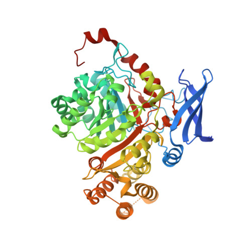
    Archaea synthesize isoprenoid-based ether-linked membrane lipids, which enable them to withstand extreme environmental conditions, such as high temperatures, high salinity, and low or high pH values 1-5 . In some archaea, such as Methanocaldococcus jannaschii, these lipids are further modified by forming carbon-carbon bonds between the termini of two lipid tails within one glycerophospholipid to generate the macrocyclic archaeol or forming two carbon-carbon bonds between the termini of two lipid tails from two glycerophospholipids to generate the macrocycle glycerol dibiphytanyl glycerol tetraether (GDGT) 1,2 . GDGT contains two 40-carbon lipid chains (biphytanyl chains) that span both leaflets of the membrane, providing enhanced stability to extreme conditions. How these specialized lipids are formed has puzzled scientists for decades. The reaction necessitates the coupling of two completely inert sp 3 -hybridized carbon centres, which, to our knowledge, has not been observed in nature. Here we show that the gene product of mj0619 from M. jannaschii, which encodes a radical S-adenosylmethionine enzyme, is responsible for biphytanyl chain formation during synthesis of both the macrocyclic archaeol and GDGT membrane lipids 6 . Structures of the enzyme show the presence of four metallocofactors: three [Fe 4 S 4 ] clusters and one mononuclear rubredoxin-like iron ion. In vitro mechanistic studies show that Csp 3 -Csp 3 bond formation takes place on fully saturated archaeal lipid substrates and involves an intermediate bond between the substrate carbon and a sulfur of one of the [Fe 4 S 4 ] clusters. Our results not only establish the biosynthetic route for tetraether formation but also improve the use of GDGT in GDGT-based paleoclimatology indices 7-10 .


  • Organizational Affiliation

    Department of Biochemistry and Molecular Biology, Pennsylvania State University, University Park, PA, USA.


Macromolecules
Find similar proteins by:  (by identity cutoff)  |  3D Structure
Entity ID: 1
MoleculeChains Sequence LengthOrganismDetailsImage
glycerol dibiphytanyl glycerol tetraether - macrocyclic archaeol synthase526Methanocaldococcus jannaschiiMutation(s): 0 
Gene Names: MJ0619
EC: 2.1.1
UniProt
Find proteins for Q58036 (Methanocaldococcus jannaschii (strain ATCC 43067 / DSM 2661 / JAL-1 / JCM 10045 / NBRC 100440))
Explore Q58036 
Go to UniProtKB:  Q58036
Entity Groups  
Sequence Clusters30% Identity50% Identity70% Identity90% Identity95% Identity100% Identity
UniProt GroupQ58036
Sequence Annotations
Expand
  • Reference Sequence
Small Molecules
Ligands 7 Unique
IDChains Name / Formula / InChI Key2D Diagram3D Interactions
L4P (Subject of Investigation/LOI)
Query on L4P

Download Ideal Coordinates CCD File 
J [auth A]3-[GLYCEROLYLPHOSPHONYL]-[1,2-DI-PHYTANYL]GLYCEROL
C46 H95 O8 P
AFYVWQWWQKSZEV-WGOBCRBGSA-N
L1P (Subject of Investigation/LOI)
Query on L1P

Download Ideal Coordinates CCD File 
H [auth A]3-PHOSPHORYL-[1,2-DI-PHYTANYL]GLYCEROL
C43 H89 O6 P
UKQGAMWGTOTQPC-ALOLAALWSA-N
SF4 (Subject of Investigation/LOI)
Query on SF4

Download Ideal Coordinates CCD File 
C [auth A],
D [auth A],
E [auth A]
IRON/SULFUR CLUSTER
Fe4 S4
LJBDFODJNLIPKO-UHFFFAOYSA-N
5AD (Subject of Investigation/LOI)
Query on 5AD

Download Ideal Coordinates CCD File 
F [auth A]5'-DEOXYADENOSINE
C10 H13 N5 O3
XGYIMTFOTBMPFP-KQYNXXCUSA-N
PGE
Query on PGE

Download Ideal Coordinates CCD File 
I [auth A]TRIETHYLENE GLYCOL
C6 H14 O4
ZIBGPFATKBEMQZ-UHFFFAOYSA-N
MET (Subject of Investigation/LOI)
Query on MET

Download Ideal Coordinates CCD File 
G [auth A]METHIONINE
C5 H11 N O2 S
FFEARJCKVFRZRR-BYPYZUCNSA-N
FE (Subject of Investigation/LOI)
Query on FE

Download Ideal Coordinates CCD File 
B [auth A]FE (III) ION
Fe
VTLYFUHAOXGGBS-UHFFFAOYSA-N
Experimental Data & Validation

Experimental Data

  • Method: X-RAY DIFFRACTION
  • Resolution: 2.03 Å
  • R-Value Free: 0.229 
  • R-Value Work: 0.186 
  • R-Value Observed: 0.189 
  • Space Group: P 21 21 21
Unit Cell:
Length ( Å )Angle ( ˚ )
a = 56.999α = 90
b = 77.598β = 90
c = 117.52γ = 90
Software Package:
Software NamePurpose
PHENIXrefinement
PDB_EXTRACTdata extraction
HKL-2000data reduction
HKL-2000data scaling
PHASERphasing

Structure Validation

View Full Validation Report


Ligand Structure Quality Assessment 


Entry History & Funding Information

Deposition Data


Funding OrganizationLocationGrant Number
National Institutes of Health/National Institute of General Medical Sciences (NIH/NIGMS)United States0000-0002-1234-8472
Howard Hughes Medical Institute (HHMI)United StatesSquire J. Booker

Revision History  (Full details and data files)

  • Version 1.0: 2022-08-31
    Type: Initial release
  • Version 1.1: 2022-09-14
    Changes: Database references
  • Version 1.2: 2023-10-18
    Changes: Data collection, Refinement description