7TIT

Cardiac thin filament decorated with regulatory M-domain of cardiac myosin binding protein C

  • Classification: MOTOR PROTEIN
  • Organism(s): Sus scrofa, Homo sapiens
  • Expression System: Escherichia coli
  • Mutation(s): No 

  • Deposited: 2022-01-14 Released: 2022-11-23 
  • Deposition Author(s): Risi, C.M., Galkin, V.E.
  • Funding Organization(s): National Institutes of Health/National Heart, Lung, and Blood Institute (NIH/NHLBI), National Institutes of Health/National Institute of General Medical Sciences (NIH/NIGMS)

Experimental Data Snapshot

  • Method: ELECTRON MICROSCOPY
  • Resolution: 8.00 Å
  • Aggregation State: HELICAL ARRAY 
  • Reconstruction Method: HELICAL 

wwPDB Validation   3D Report Full Report


This is version 2.0 of the entry. See complete history


Literature

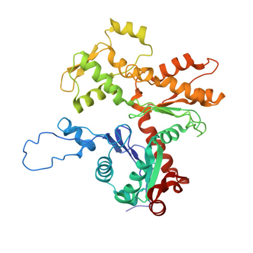
Cryo-Electron Microscopy Reveals Cardiac Myosin Binding Protein-C M-Domain Interactions with the Thin Filament.

Risi, C.M.Villanueva, E.Belknap, B.Sadler, R.L.Harris, S.P.White, H.D.Galkin, V.E.

(2022) J Mol Biol 434: 167879-167879

  • DOI: https://doi.org/10.1016/j.jmb.2022.167879
  • Primary Citation of Related Structures:  
    7TIJ, 7TIT, 7TJ7

  • PubMed Abstract: 

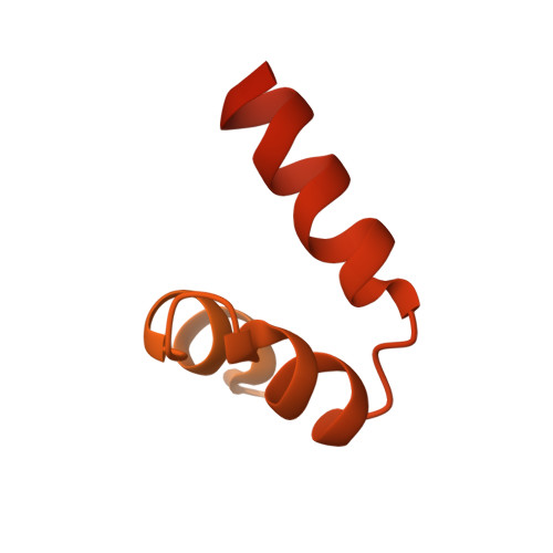
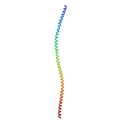
    Cardiac myosin binding protein C (cMyBP-C) modulates cardiac contraction via direct interactions with cardiac thick (myosin) and thin (actin) filaments (cTFs). While its C-terminal domains (e.g. C8-C10) anchor cMyBP-C to the backbone of the thick filament, its N-terminal domains (NTDs) (e.g. C0, C1, M, and C2) bind to both myosin and actin to accomplish its dual roles of inhibiting thick filaments and activating cTFs. While the positions of C0, C1 and C2 on cTF have been reported, the binding site of the M-domain on the surface of the cTF is unknown. Here, we used cryo-EM to reveal that the M-domain interacts with actin via helix 3 of its ordered tri-helix bundle region, while the unstructured part of the M-domain does not maintain extensive interactions with actin. We combined the recently obtained structure of the cTF with the positions of all the four NTDs on its surface to propose a complete model of the NTD binding to the cTF. The model predicts that the interactions of the NTDs with the cTF depend on the activation state of the cTF. At the peak of systole, when bound to the extensively activated cTF, NTDs would inhibit actomyosin interactions. In contrast, at falling Ca 2+ levels, NTDs would not compete with the myosin heads for binding to the cTF, but would rather promote formation of active cross-bridges at the adjacent regulatory units located at the opposite cTF strand. Our structural data provides a testable model of the cTF regulation by the cMyBP-C.


  • Organizational Affiliation

    Department of Physiological Sciences, Eastern Virginia Medical School, Norfolk, VA 23507, USA.


Macromolecules
Find similar proteins by:  (by identity cutoff)  |  3D Structure
Entity ID: 1
MoleculeChains Sequence LengthOrganismDetailsImage
cardiac actin
A, B, C, D, E
A, B, C, D, E, F
375Sus scrofaMutation(s): 0 
UniProt
Find proteins for P68137 (Sus scrofa)
Explore P68137 
Go to UniProtKB:  P68137
Entity Groups  
Sequence Clusters30% Identity50% Identity70% Identity90% Identity95% Identity100% Identity
UniProt GroupP68137
Sequence Annotations
Expand
  • Reference Sequence
Find similar proteins by:  (by identity cutoff)  |  3D Structure
Entity ID: 2
MoleculeChains Sequence LengthOrganismDetailsImage
Myosin-binding protein C, cardiac-type
G, H, I, J, K
G, H, I, J, K, L
220Homo sapiensMutation(s): 0 
Gene Names: MYBPC3
UniProt & NIH Common Fund Data Resources
Find proteins for Q14896 (Homo sapiens)
Explore Q14896 
Go to UniProtKB:  Q14896
PHAROS:  Q14896
GTEx:  ENSG00000134571 
Entity Groups  
Sequence Clusters30% Identity50% Identity70% Identity90% Identity95% Identity100% Identity
UniProt GroupQ14896
Sequence Annotations
Expand
  • Reference Sequence
Find similar proteins by:  (by identity cutoff)  |  3D Structure
Entity ID: 3
MoleculeChains Sequence LengthOrganismDetailsImage
tropomyosin model
M, N, O, P
135Sus scrofaMutation(s): 0 
Sequence Annotations
Expand
  • Reference Sequence
Experimental Data & Validation

Experimental Data

  • Method: ELECTRON MICROSCOPY
  • Resolution: 8.00 Å
  • Aggregation State: HELICAL ARRAY 
  • Reconstruction Method: HELICAL 
EM Software:
TaskSoftware PackageVersion
RECONSTRUCTIONIHRSR
MODEL REFINEMENTPHENIX

Structure Validation

View Full Validation Report


Entry History & Funding Information

Deposition Data


Funding OrganizationLocationGrant Number
National Institutes of Health/National Heart, Lung, and Blood Institute (NIH/NHLBI)United StatesHL140925
National Institutes of Health/National Institute of General Medical Sciences (NIH/NIGMS)United StatesGM116790

Revision History  (Full details and data files)

  • Version 1.0: 2022-11-23
    Type: Initial release
  • Version 1.1: 2022-12-07
    Changes: Database references, Refinement description
  • Version 2.0: 2023-03-22
    Type: Coordinate replacement
    Reason: Atoms with unrealistic or zero occupancies
    Changes: Advisory, Atomic model, Data collection, Source and taxonomy, Structure summary