Toxic antiphage defense proteins inhibited by intragenic antitoxin proteins.
Zhong, A., Jiang, X., Hickman, A.B., Klier, K., Teodoro, G.I.C., Dyda, F., Laub, M.T., Storz, G.(2023) Proc Natl Acad Sci U S A 120: e2307382120-e2307382120
- PubMed: 37487082 Search on PubMedSearch on PubMed Central
- DOI: https://doi.org/10.1073/pnas.2307382120
- Primary Citation Related Structures:
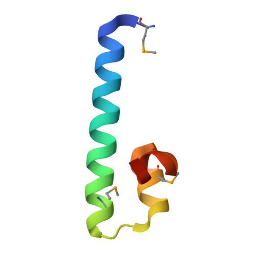
7TH0 - PubMed Abstract:
Recombination-promoting nuclease (Rpn) proteins are broadly distributed across bacterial phyla, yet their functions remain unclear. Here, we report that these proteins are toxin-antitoxin systems, comprised of genes-within-genes, that combat phage infection. We show the small, highly variable Rpn C -terminal domains (Rpn S ), which are translated separately from the full-length proteins (Rpn L ), directly block the activities of the toxic Rpn L . The crystal structure of RpnA S revealed a dimerization interface encompassing α helix that can have four amino acid repeats whose number varies widely among strains of the same species. Consistent with strong selection for the variation, we document that plasmid-encoded RpnP2 L protects Escherichia coli against certain phages. We propose that many more intragenic-encoded proteins that serve regulatory roles remain to be discovered in all organisms.
- Division of Molecular and Cellular Biology, Eunice Kennedy Shriver National Institute of Child Health and Human Development, Bethesda, MD 20892.
Organizational Affiliation: