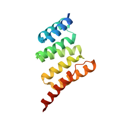
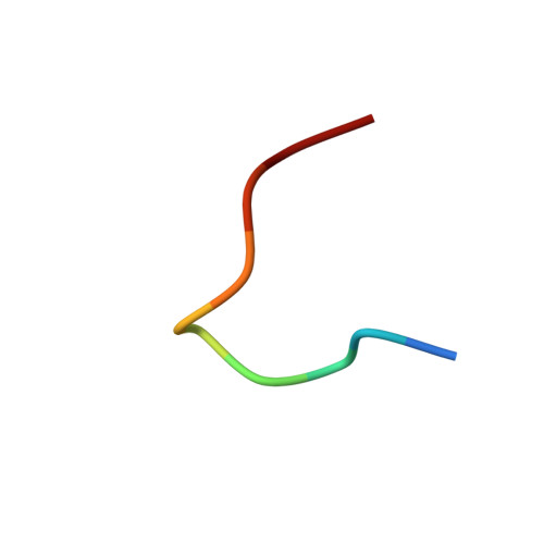
7TB1

Crystal structure of STUB1 with a macrocyclic peptide


Experimental Data Snapshot

  • Method: X-RAY DIFFRACTION
  • Resolution: 1.78 Å
  • R-Value Free: 0.278 
  • R-Value Work: 0.224 
  • R-Value Observed: 0.227 

wwPDB Validation   3D Report Full Report


Ligand Structure Quality Assessment 


This is version 1.1 of the entry. See complete history


Literature

Discovery and Structure-Based Design of Macrocyclic Peptides Targeting STUB1.

Ng, S.Brueckner, A.C.Bahmanjah, S.Deng, Q.Johnston, J.M.Ge, L.Duggal, R.Habulihaz, B.Barlock, B.Ha, S.Sadruddin, A.Yeo, C.Strickland, C.Peier, A.Henry, B.Sherer, E.C.Partridge, A.W.

(2022) J Med Chem 

  • DOI: https://doi.org/10.1021/acs.jmedchem.2c00406
  • Primary Citation of Related Structures:  
    7TB1

  • PubMed Abstract: 

    Recent evidence suggests that deletion of STUB1─a pivotal negative regulator of interferon-γ sensing─may potentially clear malignant cells. However, current studies rely primarily on genetic approaches, as pharmacological inhibitors of STUB1 are lacking. Identifying a tool compound will be a step toward validating the target in a broader therapeutic sense. Herein, screening more than a billion macrocyclic peptides resulted in STUB1 binders, which were further optimized by a structure-enabled in silico design. The strategy to replace the macrocyclic peptides' hydrophilic and solvent-exposed region with a hydrophobic scaffold improved cellular permeability while maintaining the binding conformation. Further substitution of the permeability-limiting terminal aspartic acid with a tetrazole bioisostere retained the binding to a certain extent while improving permeability, suggesting a path forward. Although not optimal for cellular study, the current lead provides a valuable template for further development into selective tool compounds for STUB1 to enable target validation.


  • Organizational Affiliation

    Quantitative Biosciences, MSD, 8 Biomedical Grove, Singapore 138665.


Macromolecules
Find similar proteins by:  (by identity cutoff)  |  3D Structure
Entity ID: 1
MoleculeChains Sequence LengthOrganismDetailsImage
E3 ubiquitin-protein ligase CHIP
A, B
140Homo sapiensMutation(s): 0 
Gene Names: STUB1CHIPPP1131
EC: 2.3.2.27
UniProt & NIH Common Fund Data Resources
Find proteins for Q9UNE7 (Homo sapiens)
Explore Q9UNE7 
Go to UniProtKB:  Q9UNE7
PHAROS:  Q9UNE7
GTEx:  ENSG00000103266 
Entity Groups  
Sequence Clusters30% Identity50% Identity70% Identity90% Identity95% Identity100% Identity
UniProt GroupQ9UNE7
Sequence Annotations
Expand
  • Reference Sequence

Find similar proteins by:  Sequence   |   3D Structure  

Entity ID: 2
MoleculeChains Sequence LengthOrganismDetailsImage
ALA-CYS-SER-SER-ILE-TRP-CYS-PRO-ASP-GLY
C, D
10synthetic constructMutation(s): 0 
Entity Groups  
Sequence Clusters30% Identity50% Identity70% Identity90% Identity95% Identity100% Identity
Sequence Annotations
Expand
  • Reference Sequence
Experimental Data & Validation

Experimental Data

  • Method: X-RAY DIFFRACTION
  • Resolution: 1.78 Å
  • R-Value Free: 0.278 
  • R-Value Work: 0.224 
  • R-Value Observed: 0.227 
  • Space Group: P 1 21 1
Unit Cell:
Length ( Å )Angle ( ˚ )
a = 36.011α = 90
b = 77.602β = 92.42
c = 46.354γ = 90
Software Package:
Software NamePurpose
BUSTERrefinement
PDB_EXTRACTdata extraction
XDSdata reduction
Aimlessdata scaling
PHASERphasing

Structure Validation

View Full Validation Report


Ligand Structure Quality Assessment 


Entry History & Funding Information

Deposition Data


Funding OrganizationLocationGrant Number
Not funded--

Revision History  (Full details and data files)

  • Version 1.0: 2022-07-27
    Type: Initial release
  • Version 1.1: 2023-10-18
    Changes: Data collection, Refinement description