7T9R | pdb_00007t9r

Crystal structure of Crocodile defensin CpoBD13:phosphatidic acid complex


Experimental Data Snapshot

  • Method: X-RAY DIFFRACTION
  • Resolution: 1.45 Å
  • R-Value Free: 
    0.215 (Depositor), 0.220 (DCC) 
  • R-Value Work: 
    0.194 (Depositor), 0.196 (DCC) 
  • R-Value Observed: 
    0.195 (Depositor) 

Starting Model: experimental
View more details

wwPDB Validation 3D Report Full Report

Validation slider image for 7T9R

Ligand Structure Quality Assessment 


This is version 1.4 of the entry. See complete history

Literature

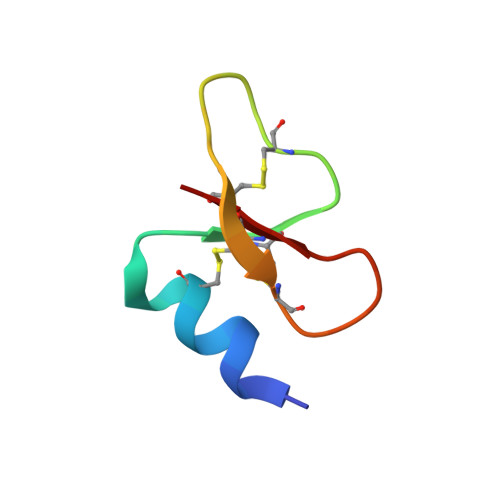
Crocodile defensin (CpoBD13) antifungal activity via pH-dependent phospholipid targeting and membrane disruption.

Williams, S.A.Lay, F.T.Bindra, G.K.Banjara, S.Poon, I.K.H.Phan, T.K.Kvansakul, M.Hulett, M.D.

(2023) Nat Commun 14: 1170-1170

  • DOI: https://doi.org/10.1038/s41467-023-36280-y
  • Primary Citation Related Structures: 
    7T9Q, 7T9R

  • PubMed Abstract: 

    Crocodilians are an order of ancient reptiles that thrive in pathogen-rich environments. The ability to inhabit these harsh environments is indicative of a resilient innate immune system. Defensins, a family of cysteine-rich cationic host defence peptides, are a major component of the innate immune systems of all plant and animal species, however crocodilian defensins are poorly characterised. We now show that the saltwater crocodile defensin CpoBD13 harbors potent antifungal activity that is mediated by a pH-dependent membrane-targeting action. CpoBD13 binds the phospholipid phosphatidic acid (PA) to form a large helical oligomeric complex, with specific histidine residues mediating PA binding. The utilisation of histidine residues for PA engagement allows CpoBD13 to exhibit differential activity at a range of environmental pH values, where CpoBD13 is optimally active in an acidic environment.


  • Organizational Affiliation
    • Department of Biochemistry and Chemistry, La Trobe Institute for Molecular Science, La Trobe University, Melbourne, 3086, Australia.

Macromolecule Content 

  • Total Structure Weight: 20.03 kDa 
  • Atom Count: 1,513 
  • Modeled Residue Count: 152 
  • Deposited Residue Count: 152 
  • Unique protein chains: 1

Macromolecules

Find similar proteins by:|  3D Structure
Entity ID: 1
MoleculeChains  Sequence LengthOrganismDetailsImage
CpoBD13
A, B, C, D
38Crocodylus porosusMutation(s): 0 
UniProt
Find proteins for A0A7G6KN55 (Crocodylus porosus)
Explore A0A7G6KN55 
Go to UniProtKB:  A0A7G6KN55
Entity Groups
Sequence Clusters30% Identity50% Identity70% Identity90% Identity95% Identity100% Identity
UniProt GroupA0A7G6KN55
Sequence Annotations
Expand
Reference Sequence

Small Molecules

Ligands 3 Unique
IDChains Name / Formula / InChI Key2D Diagram3D Interactions
PA6
(Subject of Investigation/LOI)

Query on PA6


Download:Ideal Coordinates CCD File
F [auth A],
G [auth A],
K [auth B],
L [auth C]
(R)-2-(FORMYLOXY)-3-(PHOSPHONOOXY)PROPYL PENTANOATE
C9 H17 O8 P
JDTMNMAQWVSSOO-MRVPVSSYSA-N
PO4

Query on PO4


Download:Ideal Coordinates CCD File
H [auth A],
I [auth A],
J [auth A],
N [auth C],
P [auth D]
PHOSPHATE ION
O4 P
NBIIXXVUZAFLBC-UHFFFAOYSA-K
EDO

Query on EDO


Download:Ideal Coordinates CCD File
E [auth A],
M [auth C],
O [auth D]
1,2-ETHANEDIOL
C2 H6 O2
LYCAIKOWRPUZTN-UHFFFAOYSA-N

Experimental Data & Validation

Experimental Data

  • Method: X-RAY DIFFRACTION
  • Resolution: 1.45 Å
  • R-Value Free:  0.215 (Depositor), 0.220 (DCC) 
  • R-Value Work:  0.194 (Depositor), 0.196 (DCC) 
  • R-Value Observed: 0.195 (Depositor) 
Space Group: P 1
Unit Cell:
Length ( Å )Angle ( ˚ )
a = 27.056α = 90.093
b = 33.694β = 90.016
c = 36.642γ = 90.014
Software Package:
Software NamePurpose
PHENIXrefinement
XDSdata reduction
Aimlessdata scaling
PHASERphasing

Structure Validation

View Full Validation Report


Ligand Structure Quality Assessment 


Entry History 

& Funding Information

Deposition Data


Funding OrganizationLocationGrant Number
Australian Research Council (ARC)AustraliaDP190103591

Revision History  (Full details and data files)

  • Version 1.0: 2023-01-18
    Type: Initial release
  • Version 1.1: 2023-02-01
    Changes: Source and taxonomy
  • Version 1.2: 2023-08-30
    Changes: Data collection, Database references
  • Version 1.3: 2023-10-25
    Changes: Refinement description
  • Version 1.4: 2024-11-13
    Changes: Structure summary