


Crystal Structure of Acyl-CoA dehydrogenase from Mycobacterium marinum in complex with FDA
DeBouver, N.D., Abendroth, J., Sroge, C.D., Lorimer, D.D., Horanyi, P.S., Edwards, T.E.To be published.
Experimental Data Snapshot
Starting Model: experimental
View more details
Entity ID: 1 | |||||
|---|---|---|---|---|---|
| Molecule | Chains | Sequence Length | Organism | Details | Image |
| Acyl-CoA dehydrogenase | 388 | Mycobacterium marinum | Mutation(s): 1 Gene Names: mmgC_16, DAVIS_02721 EC: 1.3.99 | 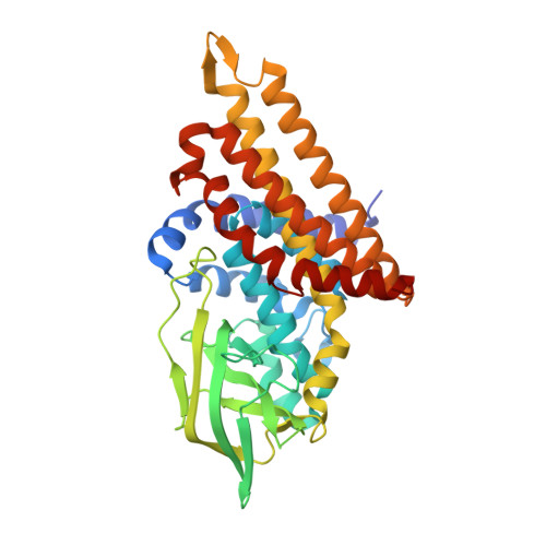 | |
UniProt | |||||
Entity Groups | |||||
| Sequence Clusters | 30% Identity50% Identity70% Identity90% Identity95% Identity100% Identity | ||||
| UniProt Group | A0A3E2MWC7 | ||||
Sequence AnnotationsExpand | |||||
Reference Sequence | |||||
| Ligands 1 Unique | |||||
|---|---|---|---|---|---|
| ID | Chains | Name / Formula / InChI Key | 2D Diagram | 3D Interactions | |
| FDA (Subject of Investigation/LOI) Download:Ideal Coordinates CCD File | C [auth A], D [auth B] | DIHYDROFLAVINE-ADENINE DINUCLEOTIDE C27 H35 N9 O15 P2 YPZRHBJKEMOYQH-UYBVJOGSSA-N |  | ||
| Length ( Å ) | Angle ( ˚ ) |
|---|---|
| a = 120.14 | α = 90 |
| b = 120.14 | β = 90 |
| c = 93.96 | γ = 120 |
| Software Name | Purpose |
|---|---|
| PHENIX | refinement |
| XDS | data reduction |
| XSCALE | data scaling |
| PDB_EXTRACT | data extraction |
| MoRDa | phasing |
| Funding Organization | Location | Grant Number |
|---|---|---|
| Not funded | -- |


