


Crystal Structure of Probable GTP-binding protein EngB bound to GDP from Klebsiella pneumoniae subsp. pneumoniae
DeBouver, N.D., Lorimer, D.D., Horanyi, P.S., Edwards, T.E.To be published.
Experimental Data Snapshot
Starting Model: experimental
View more details
Entity ID: 1 | |||||
|---|---|---|---|---|---|
| Molecule | Chains | Sequence Length | Organism | Details | Image |
| Probable GTP-binding protein EngB | 219 | Klebsiella pneumoniae subsp. pneumoniae HS11286 | Mutation(s): 0 Gene Names: engB, KPHS_00220 | 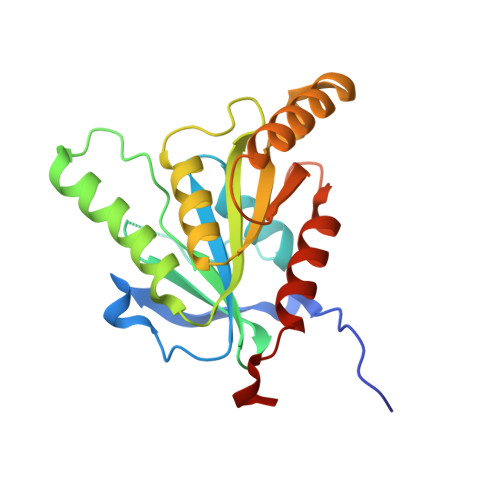 | |
UniProt | |||||
Entity Groups | |||||
| Sequence Clusters | 30% Identity50% Identity70% Identity90% Identity95% Identity100% Identity | ||||
| UniProt Group | A6TG68 | ||||
Sequence AnnotationsExpand | |||||
Reference Sequence | |||||
| Ligands 4 Unique | |||||
|---|---|---|---|---|---|
| ID | Chains | Name / Formula / InChI Key | 2D Diagram | 3D Interactions | |
| GDP (Subject of Investigation/LOI) Download:Ideal Coordinates CCD File | C [auth A], E [auth B] | GUANOSINE-5'-DIPHOSPHATE C10 H15 N5 O11 P2 QGWNDRXFNXRZMB-UUOKFMHZSA-N |  | ||
| GOL Download:Ideal Coordinates CCD File | D [auth A] | GLYCEROL C3 H8 O3 PEDCQBHIVMGVHV-UHFFFAOYSA-N |  | ||
| BU1 Download:Ideal Coordinates CCD File | F [auth B] | 1,4-BUTANEDIOL C4 H10 O2 WERYXYBDKMZEQL-UHFFFAOYSA-N |  | ||
| PGR Download:Ideal Coordinates CCD File | G [auth B] | R-1,2-PROPANEDIOL C3 H8 O2 DNIAPMSPPWPWGF-GSVOUGTGSA-N |  | ||
| Length ( Å ) | Angle ( ˚ ) |
|---|---|
| a = 105.47 | α = 90 |
| b = 94.07 | β = 108.6 |
| c = 56.59 | γ = 90 |
| Software Name | Purpose |
|---|---|
| XDS | data reduction |
| XSCALE | data scaling |
| PHENIX | refinement |
| PDB_EXTRACT | data extraction |
| MoRDa | phasing |
| Funding Organization | Location | Grant Number |
|---|---|---|
| Not funded | -- |


