7SX8

T-Plastin-F-actin complex, parallel bundled state


Experimental Data Snapshot

  • Method: ELECTRON MICROSCOPY
  • Resolution: 9.00 Å
  • Aggregation State: FILAMENT 
  • Reconstruction Method: SINGLE PARTICLE 

wwPDB Validation   3D Report Full Report


This is version 1.1 of the entry. See complete history


Literature

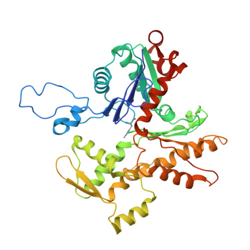
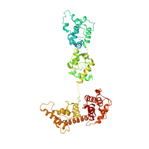
Structural mechanism for bidirectional actin cross-linking by T-plastin.

Mei, L.Reynolds, M.J.Garbett, D.Gong, R.Meyer, T.Alushin, G.M.

(2022) Proc Natl Acad Sci U S A 119: e2205370119-e2205370119

  • DOI: https://doi.org/10.1073/pnas.2205370119
  • Primary Citation of Related Structures:  
    7R94, 7SX8, 7SX9, 7SXA

  • PubMed Abstract: 

    To orchestrate cell mechanics, trafficking, and motility, cytoskeletal filaments must assemble into higher-order networks whose local subcellular architecture and composition specify their functions. Cross-linking proteins bridge filaments at the nanoscale to control a network's μm-scale geometry, thereby conferring its mechanical properties and functional dynamics. While these interfilament linkages are key determinants of cytoskeletal function, their structural mechanisms remain poorly understood. Plastins/fimbrins are an evolutionarily ancient family of tandem calponin-homology domain (CHD) proteins required to construct multiple classes of actin networks, which feature diverse geometries specialized to power cytokinesis, microvilli and stereocilia biogenesis, and persistent cell migration. Here, we focus on the structural basis of actin network assembly by human T-plastin, a ubiquitously expressed isoform necessary for the maintenance of stable cellular protrusions generated by actin polymerization forces. By implementing a machine-learning-enabled cryo-electron microscopy pipeline for visualizing cross-linkers bridging multiple filaments, we uncover a sequential bundling mechanism enabling T-plastin to bridge pairs of actin filaments in both parallel and antiparallel orientations. T-plastin populates distinct structural landscapes in these two bridging orientations that are selectively compatible with actin networks featuring divergent architectures and functions. Our structural, biochemical, and cell biological data highlight inter-CHD linkers as key structural elements underlying flexible but stable cross-linking that are likely to be disrupted by T-plastin mutations that cause hereditary bone diseases.


  • Organizational Affiliation

    Laboratory of Structural Biophysics and Mechanobiology, The Rockefeller University, New York, NY 10065.


Macromolecules
Find similar proteins by:  (by identity cutoff)  |  3D Structure
Entity ID: 1
MoleculeChains Sequence LengthOrganismDetailsImage
Actin, alpha skeletal muscle377Gallus gallusMutation(s): 0 
UniProt
Find proteins for P68139 (Gallus gallus)
Explore P68139 
Go to UniProtKB:  P68139
Entity Groups  
Sequence Clusters30% Identity50% Identity70% Identity90% Identity95% Identity100% Identity
UniProt GroupP68139
Sequence Annotations
Expand
  • Reference Sequence
Find similar proteins by:  (by identity cutoff)  |  3D Structure
Entity ID: 2
MoleculeChains Sequence LengthOrganismDetailsImage
Plastin-3630Homo sapiensMutation(s): 0 
Gene Names: PLS3
UniProt & NIH Common Fund Data Resources
Find proteins for P13797 (Homo sapiens)
Explore P13797 
Go to UniProtKB:  P13797
PHAROS:  P13797
GTEx:  ENSG00000102024 
Entity Groups  
Sequence Clusters30% Identity50% Identity70% Identity90% Identity95% Identity100% Identity
UniProt GroupP13797
Sequence Annotations
Expand
  • Reference Sequence
Small Molecules
Ligands 2 Unique
IDChains Name / Formula / InChI Key2D Diagram3D Interactions
ADP
Query on ADP

Download Ideal Coordinates CCD File 
H [auth C]
J [auth B]
L [auth A]
N [auth H]
P [auth G]
H [auth C],
J [auth B],
L [auth A],
N [auth H],
P [auth G],
R [auth E]
ADENOSINE-5'-DIPHOSPHATE
C10 H15 N5 O10 P2
XTWYTFMLZFPYCI-KQYNXXCUSA-N
MG
Query on MG

Download Ideal Coordinates CCD File 
I [auth C]
K [auth B]
M [auth A]
O [auth H]
Q [auth G]
I [auth C],
K [auth B],
M [auth A],
O [auth H],
Q [auth G],
S [auth E]
MAGNESIUM ION
Mg
JLVVSXFLKOJNIY-UHFFFAOYSA-N
Modified Residues  1 Unique
IDChains TypeFormula2D DiagramParent
HIC
Query on HIC
A [auth C]
B
C [auth A]
E [auth H]
F [auth G]
A [auth C],
B,
C [auth A],
E [auth H],
F [auth G],
G [auth E]
L-PEPTIDE LINKINGC7 H11 N3 O2HIS
Experimental Data & Validation

Experimental Data

  • Method: ELECTRON MICROSCOPY
  • Resolution: 9.00 Å
  • Aggregation State: FILAMENT 
  • Reconstruction Method: SINGLE PARTICLE 
EM Software:
TaskSoftware PackageVersion
MODEL REFINEMENTPHENIX
RECONSTRUCTIONRELION3.1

Structure Validation

View Full Validation Report


Entry History & Funding Information

Deposition Data


Funding OrganizationLocationGrant Number
National Institutes of Health/National Institute of Dental and Craniofacial Research (NIH/NIDCR)United States5DP5OD017885
National Institutes of Health/National Institute of General Medical Sciences (NIH/NIGMS)United StatesR01GM141044

Revision History  (Full details and data files)

  • Version 1.0: 2022-08-31
    Type: Initial release
  • Version 1.1: 2022-09-21
    Changes: Database references