7SUC

XFEL Serial Crystallography Reveals the Room Temperature Structure of Methyl-Coenzyme M Reductase


Experimental Data Snapshot

  • Method: X-RAY DIFFRACTION
  • Resolution: 1.90 Å
  • R-Value Free: 0.186 
  • R-Value Work: 0.152 
  • R-Value Observed: 0.153 

wwPDB Validation   3D Report Full Report


Ligand Structure Quality Assessment 


This is version 1.3 of the entry. See complete history


Literature

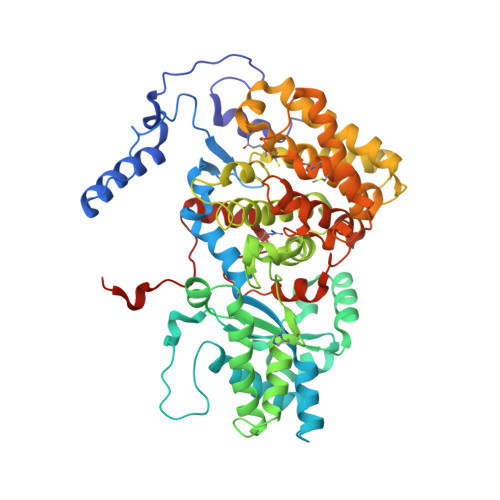
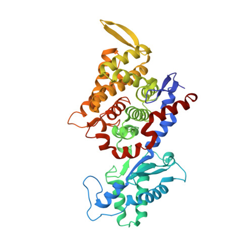
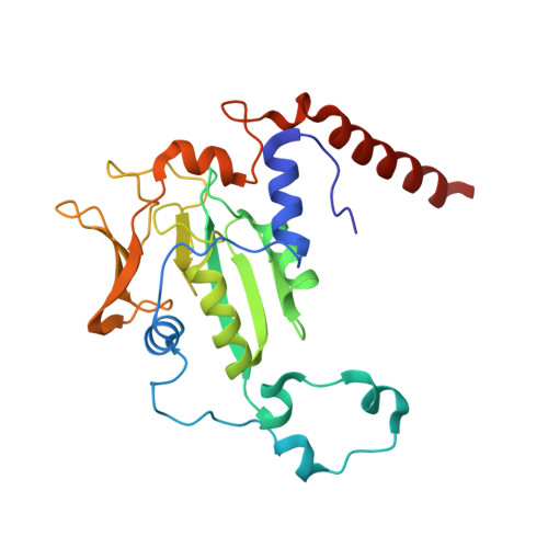
XFEL serial crystallography reveals the room temperature structure of methyl-coenzyme M reductase.

Ohmer, C.J.Dasgupta, M.Patwardhan, A.Bogacz, I.Kaminsky, C.Doyle, M.D.Chen, P.Y.Keable, S.M.Makita, H.Simon, P.S.Massad, R.Fransson, T.Chatterjee, R.Bhowmick, A.Paley, D.W.Moriarty, N.W.Brewster, A.S.Gee, L.B.Alonso-Mori, R.Moss, F.Fuller, F.D.Batyuk, A.Sauter, N.K.Bergmann, U.Drennan, C.L.Yachandra, V.K.Yano, J.Kern, J.F.Ragsdale, S.W.

(2022) J Inorg Biochem 230: 111768-111768

  • DOI: https://doi.org/10.1016/j.jinorgbio.2022.111768
  • Primary Citation of Related Structures:  
    7SUC, 7SXM

  • PubMed Abstract: 

    Methyl-Coenzyme M Reductase (MCR) catalyzes the biosynthesis of methane in methanogenic archaea, using a catalytic Ni-centered Cofactor F430 in its active site. It also catalyzes the reverse reaction, that is, the anaerobic activation and oxidation, including the cleavage of the CH bond in methane. Because methanogenesis is the major source of methane on earth, understanding the reaction mechanism of this enzyme can have massive implications in global energy balances. While recent publications have proposed a radical-based catalytic mechanism as well as novel sulfonate-based binding modes of MCR for its native substrates, the structure of the active state of MCR, as well as a complete characterization of the reaction, remain elusive. Previous attempts to structurally characterize the active MCR-Ni(I) state have been unsuccessful due to oxidation of the redox- sensitive catalytic Ni center. Further, while many cryo structures of the inactive Ni(II)-enzyme in various substrates-bound forms have been published, no room temperature structures have been reported, and the structure and mechanism of MCR under physiologically relevant conditions is not known. In this study, we report the first room temperature structure of the MCRred1-silent Ni(II) form using an X-ray Free-Electron Laser (XFEL), with simultaneous X-ray Emission Spectroscopy (XES) and X-ray Diffraction (XRD) data collection. In celebration of the seminal contributions of inorganic chemist Dick Holm to our understanding of nickel-based catalysis, we are honored to announce our findings in this special issue dedicated to this remarkable pioneer of bioinorganic chemistry.


  • Organizational Affiliation

    Department of Biological Chemistry, University of Michigan Medical School, 1150 W. Medical Center Dr., 5200 MSRBIII, Ann Arbor, MI 48109-0606, USA.


Macromolecules
Find similar proteins by:  (by identity cutoff)  |  3D Structure
Entity ID: 1
MoleculeChains Sequence LengthOrganismDetailsImage
Methyl-coenzyme M reductase I subunit alphaA,
D [auth a]
548Methanothermobacter marburgensis str. MarburgMutation(s): 0 
EC: 2.8.4.1
UniProt
Find proteins for P11558 (Methanothermobacter marburgensis (strain ATCC BAA-927 / DSM 2133 / JCM 14651 / NBRC 100331 / OCM 82 / Marburg))
Explore P11558 
Go to UniProtKB:  P11558
Entity Groups  
Sequence Clusters30% Identity50% Identity70% Identity90% Identity95% Identity100% Identity
UniProt GroupP11558
Sequence Annotations
Expand
  • Reference Sequence
Find similar proteins by:  (by identity cutoff)  |  3D Structure
Entity ID: 2
MoleculeChains Sequence LengthOrganismDetailsImage
Methyl-coenzyme M reductase I subunit betaB,
E [auth b]
442Methanothermobacter marburgensis str. MarburgMutation(s): 0 
EC: 2.8.4.1
UniProt
Find proteins for P11560 (Methanothermobacter marburgensis (strain ATCC BAA-927 / DSM 2133 / JCM 14651 / NBRC 100331 / OCM 82 / Marburg))
Explore P11560 
Go to UniProtKB:  P11560
Entity Groups  
Sequence Clusters30% Identity50% Identity70% Identity90% Identity95% Identity100% Identity
UniProt GroupP11560
Sequence Annotations
Expand
  • Reference Sequence
Find similar proteins by:  (by identity cutoff)  |  3D Structure
Entity ID: 3
MoleculeChains Sequence LengthOrganismDetailsImage
Methyl-coenzyme M reductase I subunit gammaC,
F [auth c]
246Methanothermobacter marburgensis str. MarburgMutation(s): 0 
EC: 2.8.4.1
UniProt
Find proteins for P11562 (Methanothermobacter marburgensis (strain ATCC BAA-927 / DSM 2133 / JCM 14651 / NBRC 100331 / OCM 82 / Marburg))
Explore P11562 
Go to UniProtKB:  P11562
Entity Groups  
Sequence Clusters30% Identity50% Identity70% Identity90% Identity95% Identity100% Identity
UniProt GroupP11562
Sequence Annotations
Expand
  • Reference Sequence
Small Molecules
Ligands 6 Unique
IDChains Name / Formula / InChI Key2D Diagram3D Interactions
F43 (Subject of Investigation/LOI)
Query on F43

Download Ideal Coordinates CCD File 
H [auth A],
Q [auth a]
FACTOR 430
C42 H51 N6 Ni O13
XLFIRMYGVLUNOY-SXMZNAGASA-M
TP7 (Subject of Investigation/LOI)
Query on TP7

Download Ideal Coordinates CCD File 
I [auth A],
L [auth A]
Coenzyme B
C11 H22 N O7 P S
JBJSVEVEEGOEBZ-SCZZXKLOSA-N
COM
Query on COM

Download Ideal Coordinates CCD File 
J [auth A],
R [auth a]
1-THIOETHANESULFONIC ACID
C2 H6 O3 S2
ZNEWHQLOPFWXOF-UHFFFAOYSA-N
EDO
Query on EDO

Download Ideal Coordinates CCD File 
S [auth a]1,2-ETHANEDIOL
C2 H6 O2
LYCAIKOWRPUZTN-UHFFFAOYSA-N
ACT
Query on ACT

Download Ideal Coordinates CCD File 
K [auth A]ACETATE ION
C2 H3 O2
QTBSBXVTEAMEQO-UHFFFAOYSA-M
MG
Query on MG

Download Ideal Coordinates CCD File 
G [auth A]
M [auth B]
N [auth C]
O [auth a]
P [auth a]
G [auth A],
M [auth B],
N [auth C],
O [auth a],
P [auth a],
T [auth b],
U [auth c]
MAGNESIUM ION
Mg
JLVVSXFLKOJNIY-UHFFFAOYSA-N
Modified Residues  6 Unique
IDChains TypeFormula2D DiagramParent
AGM
Query on AGM
A,
D [auth a]
L-PEPTIDE LINKINGC7 H17 N4 O2ARG
DYA
Query on DYA
A,
D [auth a]
L-PEPTIDE LINKINGC4 H5 N O4ASP
GL3
Query on GL3
A,
D [auth a]
L-PEPTIDE LINKINGC2 H5 N O SGLY
MGN
Query on MGN
A,
D [auth a]
L-PEPTIDE LINKINGC6 H12 N2 O3GLN
MHS
Query on MHS
A,
D [auth a]
L-PEPTIDE LINKINGC7 H11 N3 O2HIS
SMC
Query on SMC
A,
D [auth a]
L-PEPTIDE LINKINGC4 H9 N O2 SCYS
Experimental Data & Validation

Experimental Data

  • Method: X-RAY DIFFRACTION
  • Resolution: 1.90 Å
  • R-Value Free: 0.186 
  • R-Value Work: 0.152 
  • R-Value Observed: 0.153 
  • Space Group: P 1 21 1
Unit Cell:
Length ( Å )Angle ( ˚ )
a = 83.082α = 90
b = 119.777β = 91.73
c = 123.21γ = 90
Software Package:
Software NamePurpose
PHENIXrefinement
cctbx.xfeldata reduction
cxi.mergedata scaling
Cootmodel building
PHASERphasing

Structure Validation

View Full Validation Report


Ligand Structure Quality Assessment 


Entry History & Funding Information

Deposition Data


Funding OrganizationLocationGrant Number
National Institutes of Health/National Institute of General Medical Sciences (NIH/NIGMS)United States1P41GM139687
National Institutes of Health/National Institute of General Medical Sciences (NIH/NIGMS)United StatesGM55302
National Institutes of Health/National Institute of General Medical Sciences (NIH/NIGMS)United StatesGM110501
National Institutes of Health/National Institute of General Medical Sciences (NIH/NIGMS)United StatesGM126289
National Institutes of Health/National Institute of General Medical Sciences (NIH/NIGMS)United StatesGM117126
National Institutes of Health/National Institute of General Medical Sciences (NIH/NIGMS)United StatesR35 GM126982
National Institutes of Health/National Institute of General Medical Sciences (NIH/NIGMS)United States--
Department of Energy (DOE, United States)United StatesDE-AC02-05CH11231
National Institutes of Health/National Institute of General Medical Sciences (NIH/NIGMS)United StatesP30 GM124169-01

Revision History  (Full details and data files)

  • Version 1.0: 2022-03-02
    Type: Initial release
  • Version 1.1: 2022-03-23
    Changes: Database references
  • Version 1.2: 2023-10-18
    Changes: Data collection, Refinement description
  • Version 1.3: 2023-11-15
    Changes: Data collection, Derived calculations