7ST8

Crystal structure of 7H2.2 Fab in complex with SAS1B C-terminal region


Experimental Data Snapshot

  • Method: X-RAY DIFFRACTION
  • Resolution: 2.75 Å
  • R-Value Free: 0.270 
  • R-Value Work: 0.231 
  • R-Value Observed: 0.233 

wwPDB Validation   3D Report Full Report


This is version 1.1 of the entry. See complete history


Literature

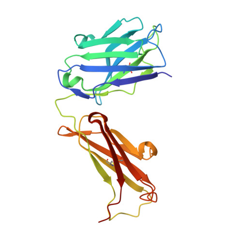
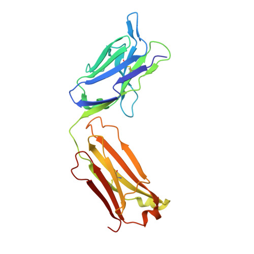
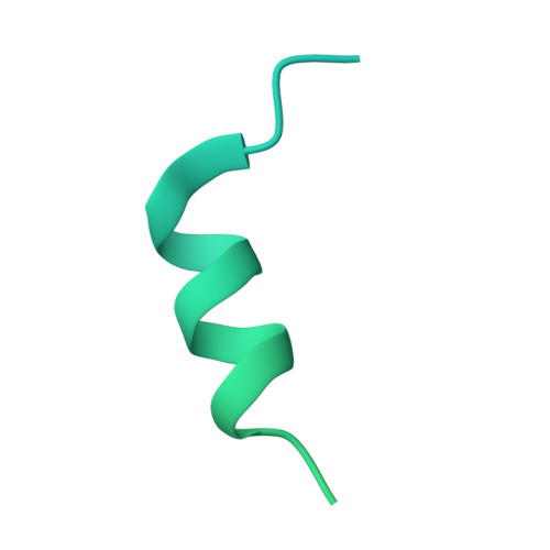
Monoclonal antibody 7H2.2 binds the C-terminus of the cancer-oocyte antigen SAS1B through the hydrophilic face of a conserved amphipathic helix corresponding to one of only two regions predicted to be ordered

Legg, M.S.G.Gagnon, S.M.L.Powell, C.J.Boulanger, M.J.Li, A.J.J.Evans, S.V.

(2022) Acta Crystallogr D Biol Crystallogr 78: 623-632

  • DOI: https://doi.org/10.1107/S2059798322003011
  • Primary Citation of Related Structures:  
    7ST8

  • PubMed Abstract: 

    The structure of the antigen-binding fragment (Fab) of mouse monoclonal antibody 7H2.2 in complex with a 15-residue fragment from the metalloproteinase sperm acrosomal SLLP1 binding protein (SAS1B), which is a molecular and cellular candidate for both cancer therapy and female contraception, has been determined at 2.75 Å resolution by single-crystal X-ray diffraction. Although the crystallization conditions contained the final 148 C-terminal residues of SAS1B, the Fab was observed to crystallize in complex with a 15-residue fragment corresponding to one of only two elements of secondary structure that are predicted to be ordered within the C-terminal region of SAS1B. The antigen forms an amphipathic α-helix that binds the 7H2.2 combining site via hydrophilic residues in an epitope that spans the length of the antigen α-helix, with only two CH-π interactions observed along the edge of the interface between the antibody and antigen. Interestingly, the paratope contains two residues mutated away from the germline (YL32F and YH58R), as well as a ProH96-ThrH97-AspH98-AspH99 insertion within heavy chain CDR3. The intact 7H2.2 antibody exhibits high affinity for the SAS1B antigen, with 1:1 binding and nanomolar affinity for both the SAS1B C-terminal construct used for crystallization (3.38 ± 0.59 nM) and a 15-amino-acid synthetic peptide construct corresponding to the helical antigen observed within the crystal structure (1.60 ± 0.31 nM). The SAS1B-antibody structure provides the first structural insight into any portion of the subdomain architecture of the C-terminal region of the novel cancer-oocyte tumor surface neoantigen SAS1B and provides a basis for the targeted use of SAS1B.


  • Organizational Affiliation

    Department of Biochemistry and Microbiology, University of Victoria, PO Box 3055 STN CSC, Victoria, BC V8Z 7X8, Canada.


Macromolecules
Find similar proteins by:  (by identity cutoff)  |  3D Structure
Entity ID: 1
MoleculeChains Sequence LengthOrganismDetailsImage
7H2.2 Fab Heavy ChainA [auth H]223Mus musculusMutation(s): 0 
Entity Groups  
Sequence Clusters30% Identity50% Identity70% Identity90% Identity95% Identity100% Identity
Sequence Annotations
Expand
  • Reference Sequence
Find similar proteins by:  (by identity cutoff)  |  3D Structure
Entity ID: 2
MoleculeChains Sequence LengthOrganismDetailsImage
7H2.2 Fab Light ChainB [auth L]217Mus musculusMutation(s): 0 
Entity Groups  
Sequence Clusters30% Identity50% Identity70% Identity90% Identity95% Identity100% Identity
Sequence Annotations
Expand
  • Reference Sequence
Find similar proteins by:  (by identity cutoff)  |  3D Structure
Entity ID: 3
MoleculeChains Sequence LengthOrganismDetailsImage
Astacin-like metalloendopeptidaseC [auth S]170Homo sapiensMutation(s): 0 
Gene Names: ASTL
EC: 3.4
UniProt & NIH Common Fund Data Resources
Find proteins for Q6HA08 (Homo sapiens)
Explore Q6HA08 
Go to UniProtKB:  Q6HA08
GTEx:  ENSG00000188886 
Entity Groups  
Sequence Clusters30% Identity50% Identity70% Identity90% Identity95% Identity100% Identity
UniProt GroupQ6HA08
Sequence Annotations
Expand
  • Reference Sequence
Experimental Data & Validation

Experimental Data

  • Method: X-RAY DIFFRACTION
  • Resolution: 2.75 Å
  • R-Value Free: 0.270 
  • R-Value Work: 0.231 
  • R-Value Observed: 0.233 
  • Space Group: C 1 2 1
Unit Cell:
Length ( Å )Angle ( ˚ )
a = 132.355α = 90
b = 43.03β = 123.89
c = 106.227γ = 90
Software Package:
Software NamePurpose
REFMACrefinement
HKL-2000data scaling
PDB_EXTRACTdata extraction
HKL-2000data reduction
PHASERphasing

Structure Validation

View Full Validation Report


Entry History & Funding Information

Deposition Data


Funding OrganizationLocationGrant Number
Natural Sciences and Engineering Research Council (NSERC, Canada)Canada499037-16

Revision History  (Full details and data files)

  • Version 1.0: 2022-05-11
    Type: Initial release
  • Version 1.1: 2023-10-18
    Changes: Data collection, Refinement description