Structure-affinity relationships of reversible proline analog inhibitors targeting proline dehydrogenase.
Bogner, A.N., Tanner, J.J.(2022) Org Biomol Chem 20: 895-905
- PubMed: 35018940 Search on PubMedSearch on PubMed Central
- DOI: https://doi.org/10.1039/d1ob02328d
- Primary Citation Related Structures:
7MWT, 7MWU, 7MWV, 7SQN - PubMed Abstract:
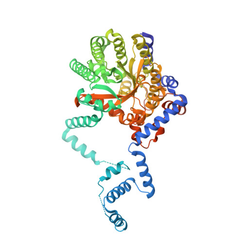
Proline dehydrogenase (PRODH) catalyzes the first step of proline catabolism, the FAD-dependent oxidation of L-proline to Δ 1 -pyrroline-5-carboxylate. PRODH plays a central role in the metabolic rewiring of cancer cells, which has motivated the discovery of inhibitors. Here, we studied the inhibition of PRODH by 18 proline-like compounds to understand the structural and chemical features responsible for the affinity of the best-known inhibitor, S -(-)-tetrahydro-2-furoic acid (1). The compounds were screened, and then six were selected for more thorough kinetic analysis: cyclobutane-1,1-dicarboxylic acid (2), cyclobutanecarboxylic acid (3), cyclopropanecarboxylic acid (4), cyclopentanecarboxylic acid (16), 2-oxobutyric acid (17), and (2 S )-oxetane-2-carboxylic acid (18). These compounds are competitive inhibitors with inhibition constants in the range of 1.4-6 mM, compared to 0.3 mM for 1. Crystal structures of PRODH complexed with 2, 3, 4, and 18 were determined. All four inhibitors bind in the proline substrate site, but the orientations of their rings differ from that of 1. The binding of 3 and 18 is accompanied by compression of the active site to enable nonpolar contacts with Leu513. Compound 2 is unique in that the additional carboxylate displaces a structurally conserved water molecule from the active site. Compound 18 also destabilizes the conserved water, but by an unexpected non-steric mechanism. The results are interpreted using a chemical double mutant thermodynamic cycle. This analysis revealed unanticipated synergism between ring size and hydrogen bonding to the conserved water. These structure-affinity relationships provide new information relevant to the development of new inhibitor design strategies targeting PRODH.
- Department of Biochemistry, University of Missouri, Columbia, Missouri 65211, USA. tannerjj@missouri.edu.
Organizational Affiliation: