7SMS

Cryo-EM structure of Torpedo acetylcholine receptor in complex with d-tubocurarine


Experimental Data Snapshot

  • Method: ELECTRON MICROSCOPY
  • Resolution: 3.18 Å
  • Aggregation State: PARTICLE 
  • Reconstruction Method: SINGLE PARTICLE 

wwPDB Validation   3D Report Full Report


This is version 1.2 of the entry. See complete history


Literature

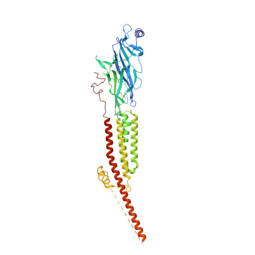
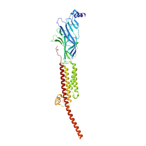
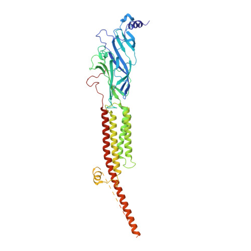
Structural mechanism of muscle nicotinic receptor desensitization and block by curare.

Rahman, M.M.Basta, T.Teng, J.Lee, M.Worrell, B.T.Stowell, M.H.B.Hibbs, R.E.

(2022) Nat Struct Mol Biol 29: 386-394

  • DOI: https://doi.org/10.1038/s41594-022-00737-3
  • Primary Citation of Related Structures:  
    7SMM, 7SMQ, 7SMR, 7SMS, 7SMT

  • PubMed Abstract: 

    Binding of the neurotransmitter acetylcholine to its receptors on muscle fibers depolarizes the membrane and thereby triggers muscle contraction. We sought to understand at the level of three-dimensional structure how agonists and antagonists alter nicotinic acetylcholine receptor conformation. We used the muscle-type receptor from the Torpedo ray to first define the structure of the receptor in a resting, activatable state. We then determined the receptor structure bound to the agonist carbachol, which stabilizes an asymmetric, closed channel desensitized state. We find conformational changes in a peripheral membrane helix are tied to recovery from desensitization. To probe mechanisms of antagonism, we obtained receptor structures with the active component of curare, a poison arrow toxin and precursor to modern muscle relaxants. d-Tubocurarine stabilizes the receptor in a desensitized-like state in the presence and absence of agonist. These findings define the transitions between resting and desensitized states and reveal divergent means by which antagonists block channel activity of the muscle-type nicotinic receptor.


  • Organizational Affiliation

    Department of Neuroscience, University of Texas Southwestern Medical Center, Dallas, TX, USA.


Macromolecules
Find similar proteins by:  (by identity cutoff)  |  3D Structure
Entity ID: 1
MoleculeChains Sequence LengthOrganismDetailsImage
Acetylcholine receptor subunit alpha
A, D
437Tetronarce californicaMutation(s): 0 
Membrane Entity: Yes 
UniProt
Find proteins for P02710 (Tetronarce californica)
Explore P02710 
Go to UniProtKB:  P02710
Entity Groups  
Sequence Clusters30% Identity50% Identity70% Identity90% Identity95% Identity100% Identity
UniProt GroupP02710
Sequence Annotations
Expand
  • Reference Sequence
Find similar proteins by:  (by identity cutoff)  |  3D Structure
Entity ID: 2
MoleculeChains Sequence LengthOrganismDetailsImage
Acetylcholine receptor subunit delta501Tetronarce californicaMutation(s): 0 
Membrane Entity: Yes 
UniProt
Find proteins for P02718 (Tetronarce californica)
Explore P02718 
Go to UniProtKB:  P02718
Entity Groups  
Sequence Clusters30% Identity50% Identity70% Identity90% Identity95% Identity100% Identity
UniProt GroupP02718
Sequence Annotations
Expand
  • Reference Sequence
Find similar proteins by:  (by identity cutoff)  |  3D Structure
Entity ID: 3
MoleculeChains Sequence LengthOrganismDetailsImage
Acetylcholine receptor subunit beta469Tetronarce californicaMutation(s): 0 
Membrane Entity: Yes 
UniProt
Find proteins for P02712 (Tetronarce californica)
Explore P02712 
Go to UniProtKB:  P02712
Entity Groups  
Sequence Clusters30% Identity50% Identity70% Identity90% Identity95% Identity100% Identity
UniProt GroupP02712
Sequence Annotations
Expand
  • Reference Sequence
Find similar proteins by:  (by identity cutoff)  |  3D Structure
Entity ID: 4
MoleculeChains Sequence LengthOrganismDetailsImage
Acetylcholine receptor subunit gamma489Tetronarce californicaMutation(s): 0 
Membrane Entity: Yes 
UniProt
Find proteins for P02714 (Tetronarce californica)
Explore P02714 
Go to UniProtKB:  P02714
Entity Groups  
Sequence Clusters30% Identity50% Identity70% Identity90% Identity95% Identity100% Identity
UniProt GroupP02714
Sequence Annotations
Expand
  • Reference Sequence
Oligosaccharides

Help

Entity ID: 5
MoleculeChains Length2D Diagram Glycosylation3D Interactions
alpha-D-mannopyranose-(1-6)-alpha-D-mannopyranose-(1-6)-beta-D-mannopyranose-(1-4)-2-acetamido-2-deoxy-beta-D-glucopyranose-(1-4)-2-acetamido-2-deoxy-beta-D-glucopyranose
F, I, J
5N/AN-Glycosylation
Glycosylation Resources
GlyTouCan:  G94626GC
GlyCosmos:  G94626GC
GlyGen:  G94626GC
Entity ID: 6
MoleculeChains Length2D Diagram Glycosylation3D Interactions
2-acetamido-2-deoxy-beta-D-glucopyranose-(1-4)-2-acetamido-2-deoxy-beta-D-glucopyranose
G
2N/AN-Glycosylation
Glycosylation Resources
GlyTouCan:  G42666HT
GlyCosmos:  G42666HT
GlyGen:  G42666HT
Entity ID: 7
MoleculeChains Length2D Diagram Glycosylation3D Interactions
beta-D-mannopyranose-(1-4)-2-acetamido-2-deoxy-beta-D-glucopyranose-(1-4)-2-acetamido-2-deoxy-beta-D-glucopyranose
H
3N/AN-Glycosylation
Glycosylation Resources
GlyTouCan:  G15407YE
GlyCosmos:  G15407YE
GlyGen:  G15407YE
Small Molecules
Ligands 3 Unique
IDChains Name / Formula / InChI Key2D Diagram3D Interactions
POV
Query on POV

Download Ideal Coordinates CCD File 
N [auth B],
O [auth C],
P [auth C],
T [auth D],
V [auth E]
(2S)-3-(hexadecanoyloxy)-2-[(9Z)-octadec-9-enoyloxy]propyl 2-(trimethylammonio)ethyl phosphate
C42 H82 N O8 P
WTJKGGKOPKCXLL-PFDVCBLKSA-N
TC9 (Subject of Investigation/LOI)
Query on TC9

Download Ideal Coordinates CCD File 
L [auth B],
M [auth B],
R [auth D],
U [auth E]
D-TUBOCURARINE
C37 H42 N2 O6
JFJZZMVDLULRGK-URLMMPGGSA-P
CLR
Query on CLR

Download Ideal Coordinates CCD File 
K [auth A],
Q [auth C],
S [auth D]
CHOLESTEROL
C27 H46 O
HVYWMOMLDIMFJA-DPAQBDIFSA-N
Experimental Data & Validation

Experimental Data

  • Method: ELECTRON MICROSCOPY
  • Resolution: 3.18 Å
  • Aggregation State: PARTICLE 
  • Reconstruction Method: SINGLE PARTICLE 
EM Software:
TaskSoftware PackageVersion
RECONSTRUCTIONRELION3.1

Structure Validation

View Full Validation Report


Entry History & Funding Information

Deposition Data


Funding OrganizationLocationGrant Number
National Institutes of Health/National Institute on Drug Abuse (NIH/NIDA)United States--

Revision History  (Full details and data files)

  • Version 1.0: 2022-03-09
    Type: Initial release
  • Version 1.1: 2022-03-30
    Changes: Database references
  • Version 1.2: 2022-04-27
    Changes: Database references