7SF4 | pdb_00007sf4

M. tb EgtD in complex with imatinib


Experimental Data Snapshot

  • Method: X-RAY DIFFRACTION
  • Resolution: 2.39 Å
  • R-Value Free: 
    0.264 (Depositor), 0.262 (DCC) 
  • R-Value Work: 
    0.221 (Depositor), 0.221 (DCC) 
  • R-Value Observed: 
    0.224 (Depositor) 

Starting Model: experimental
View more details

wwPDB Validation 3D Report Full Report

Validation slider image for 7SF4

Ligand Structure Quality Assessment 


This is version 1.1 of the entry. See complete history

Literature

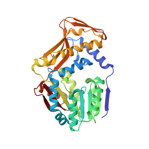
Inhibitors of Mycobacterium tuberculosis EgtD target both substrate binding sites to limit hercynine production.

Sudasinghe, T.D.Banco, M.T.Ronning, D.R.

(2021) Sci Rep 11: 22240-22240

  • DOI: https://doi.org/10.1038/s41598-021-01526-6
  • Primary Citation Related Structures: 
    7SCF, 7SEW, 7SEX, 7SEY, 7SF4, 7SF5

  • PubMed Abstract: 

    Ergothioneine (EGT) is a low molecular weight histidine betaine essential in all domains of life but only synthesized by selected few organisms. Synthesis of EGT by Mycobacterium tuberculosis (M. tb) is critical for maintaining bioenergetic homeostasis and protecting the bacterium from alkylating agents, oxidative stress, and anti-tubercular drugs. EgtD, an S-adenosylmethionine-dependent methyltransferase (AdoMet), catalyzes the trimethylation of L-Histidine to initiate EGT biosynthesis and this reaction has been shown to be essential for EGT production in mycobacteria and for long-term infection of murine macrophages by M. tb. In this work, library screening and structure-guided strategies identified multiple classes of M. tb EgtD inhibitors that bind in various regions of the enzyme active site. X-ray crystal structures of EgtD-inhibitor complexes confirm that L-Histidine analogs bind solely to the L-Histidine binding site while drug-like inhibitors, such as TGX-221, and S-Glycyl-H-1152 span both the L-Histidine and AdoMet binding sites. These enzyme-inhibitor complexes provide detailed structural information of compound scaffolds useful for developing more potent inhibitors that could shorten Tuberculosis treatment regimens by weakening important bacterial defenses.


  • Organizational Affiliation
    • Department of Pharmaceutical Sciences, University of Nebraska Medical Center, Omaha, NE, 68198, USA.

Macromolecule Content 

  • Total Structure Weight: 72.02 kDa 
  • Atom Count: 5,246 
  • Modeled Residue Count: 642 
  • Deposited Residue Count: 642 
  • Unique protein chains: 1

Macromolecules

Find similar proteins by:|  3D Structure
Entity ID: 1
MoleculeChains  Sequence LengthOrganismDetailsImage
Histidine N-alpha-methyltransferase
A, B
321Mycobacterium tuberculosisMutation(s): 0 
Gene Names: egtD
EC: 2.1.1.44
UniProt
Find proteins for P9WN47 (Mycobacterium tuberculosis (strain ATCC 25618 / H37Rv))
Explore P9WN47 
Go to UniProtKB:  P9WN47
Entity Groups
Sequence Clusters30% Identity50% Identity70% Identity90% Identity95% Identity100% Identity
UniProt GroupP9WN47
Sequence Annotations
Expand
Reference Sequence

Small Molecules

Ligands 3 Unique
IDChains Name / Formula / InChI Key2D Diagram3D Interactions
XDK
(Subject of Investigation/LOI)

Query on XDK


Download:Ideal Coordinates CCD File
C [auth A],
N [auth B]
2-AMINO-4-PYRIDYL-PYRIMIDINE
C9 H8 N4
LQHQKYWYKPLKCH-UHFFFAOYSA-N
GOL

Query on GOL


Download:Ideal Coordinates CCD File
D [auth A]
E [auth A]
F [auth A]
L [auth A]
O [auth B]
D [auth A],
E [auth A],
F [auth A],
L [auth A],
O [auth B],
Q [auth B]
GLYCEROL
C3 H8 O3
PEDCQBHIVMGVHV-UHFFFAOYSA-N
EDO

Query on EDO


Download:Ideal Coordinates CCD File
G [auth A]
H [auth A]
I [auth A]
J [auth A]
K [auth A]
G [auth A],
H [auth A],
I [auth A],
J [auth A],
K [auth A],
M [auth A],
P [auth B],
R [auth B]
1,2-ETHANEDIOL
C2 H6 O2
LYCAIKOWRPUZTN-UHFFFAOYSA-N

Experimental Data & Validation

Experimental Data

  • Method: X-RAY DIFFRACTION
  • Resolution: 2.39 Å
  • R-Value Free:  0.264 (Depositor), 0.262 (DCC) 
  • R-Value Work:  0.221 (Depositor), 0.221 (DCC) 
  • R-Value Observed: 0.224 (Depositor) 
Space Group: P 21 21 21
Unit Cell:
Length ( Å )Angle ( ˚ )
a = 68.98α = 90
b = 71.131β = 90
c = 144.557γ = 90
Software Package:
Software NamePurpose
PHENIXrefinement
HKL-2000data scaling
PDB_EXTRACTdata extraction
HKL-2000data reduction
PHASERphasing

Structure Validation

View Full Validation Report


Ligand Structure Quality Assessment 


Entry History 

& Funding Information

Deposition Data


Funding OrganizationLocationGrant Number
National Institutes of Health/National Institute Of Allergy and Infectious Diseases (NIH/NIAID)United States--

Revision History  (Full details and data files)

  • Version 1.0: 2021-12-01
    Type: Initial release
  • Version 1.1: 2023-10-18
    Changes: Data collection, Refinement description