Dynamic HIV-1 spike motion creates vulnerability for its membrane-bound tripod to antibody attack.
Yang, S., Hiotis, G., Wang, Y., Chen, J., Wang, J.H., Kim, M., Reinherz, E.L., Walz, T.(2022) Nat Commun 13: 6393-6393
- PubMed: 36302771 Search on PubMedSearch on PubMed Central
- DOI: https://doi.org/10.1038/s41467-022-34008-y
- Primary Citation Related Structures:
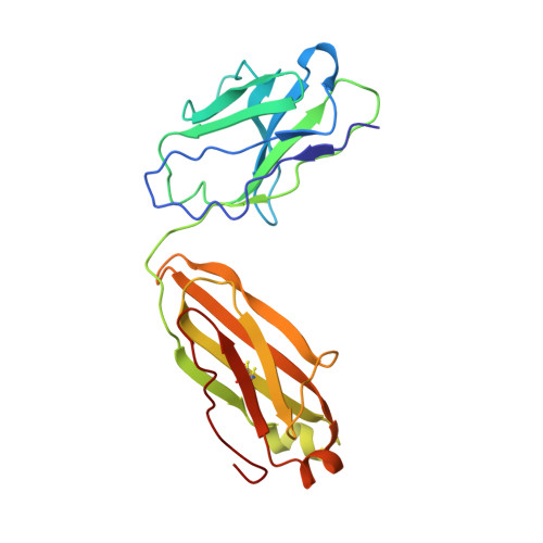
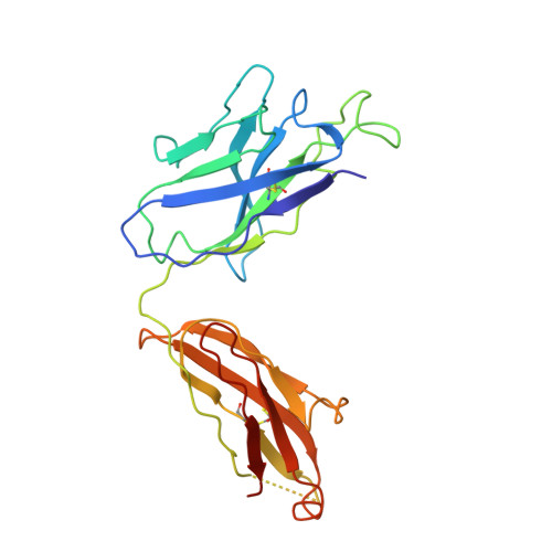
7SC5, 7SD3 - PubMed Abstract:
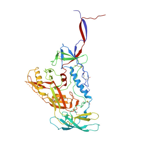
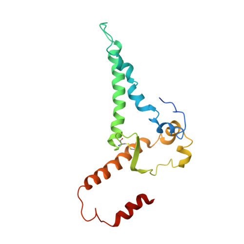
Vaccines targeting HIV-1's gp160 spike protein are stymied by high viral mutation rates and structural chicanery. gp160's membrane-proximal external region (MPER) is the target of naturally arising broadly neutralizing antibodies (bnAbs), yet MPER-based vaccines fail to generate bnAbs. Here, nanodisc-embedded spike protein was investigated by cryo-electron microscopy and molecular-dynamics simulations, revealing spontaneous ectodomain tilting that creates vulnerability for HIV-1. While each MPER protomer radiates centrally towards the three-fold axis contributing to a membrane-associated tripod structure that is occluded in the upright spike, tilting provides access to the opposing MPER. Structures of spike proteins with bound 4E10 bnAb Fabs reveal that the antibody binds exposed MPER, thereby altering MPER dynamics, modifying average ectodomain tilt, and imposing strain on the viral membrane and the spike's transmembrane segments, resulting in the abrogation of membrane fusion and informing future vaccine development.
- Laboratory of Molecular Electron Microscopy, The Rockefeller University, New York, NY, USA.
Organizational Affiliation: