7S77

Crystal structure of the G391V variant of human PGM-1


Experimental Data Snapshot

  • Method: X-RAY DIFFRACTION
  • Resolution: 2.80 Å
  • R-Value Free: 0.252 
  • R-Value Work: 0.193 
  • R-Value Observed: 0.196 

wwPDB Validation   3D Report Full Report


This is version 1.2 of the entry. See complete history


Literature

Effects of the T337M and G391V disease-related variants on human phosphoglucomutase 1: structural disruptions large and small.

Stiers, K.M.Owuocha, L.F.Beamer, L.J.

(2022) Acta Crystallogr F Struct Biol Commun 78: 200-209

  • DOI: https://doi.org/10.1107/S2053230X22004174
  • Primary Citation of Related Structures:  
    7S0W, 7S77

  • PubMed Abstract: 

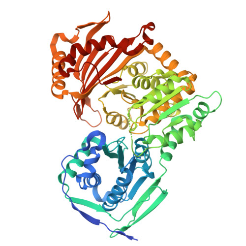
    Phosphoglucomutase 1 (PGM1) plays a central role in glucose homeostasis in human cells. Missense variants of this enzyme cause an inborn error of metabolism, which is categorized as a congenital disorder of glycosylation. Here, two disease-related variants of PGM1, T337M and G391V, which are both located in domain 3 of the four-domain protein, were characterized via X-ray crystallography and biochemical assays. The studies show multiple impacts resulting from these dysfunctional variants, including both short- and long-range structural perturbations. In the T337M variant these are limited to a small shift in an active-site loop, consistent with reduced enzyme activity. In contrast, the G391V variant produces a cascade of structural perturbations, including displacement of both the catalytic phosphoserine and metal-binding loops. This work reinforces several themes that were found in prior studies of dysfunctional PGM1 variants, including increased structural flexibility and the outsized impacts of mutations affecting interdomain interfaces. The molecular mechanisms of PGM1 variants have implications for newly described inherited disorders of related enzymes.


  • Organizational Affiliation

    Biochemistry Department, University of Missouri, Columbia, MO 65211, USA.


Macromolecules
Find similar proteins by:  (by identity cutoff)  |  3D Structure
Entity ID: 1
MoleculeChains Sequence LengthOrganismDetailsImage
Phosphoglucomutase-1
A, B
585Homo sapiensMutation(s): 1 
Gene Names: PGM1
EC: 5.4.2.2
UniProt & NIH Common Fund Data Resources
Find proteins for P36871 (Homo sapiens)
Explore P36871 
Go to UniProtKB:  P36871
PHAROS:  P36871
GTEx:  ENSG00000079739 
Entity Groups  
Sequence Clusters30% Identity50% Identity70% Identity90% Identity95% Identity100% Identity
UniProt GroupP36871
Sequence Annotations
Expand
  • Reference Sequence
Small Molecules
Ligands 1 Unique
IDChains Name / Formula / InChI Key2D Diagram3D Interactions
SO4
Query on SO4

Download Ideal Coordinates CCD File 
C [auth A]SULFATE ION
O4 S
QAOWNCQODCNURD-UHFFFAOYSA-L
Experimental Data & Validation

Experimental Data

  • Method: X-RAY DIFFRACTION
  • Resolution: 2.80 Å
  • R-Value Free: 0.252 
  • R-Value Work: 0.193 
  • R-Value Observed: 0.196 
  • Space Group: P 41 21 2
Unit Cell:
Length ( Å )Angle ( ˚ )
a = 171.05α = 90
b = 171.05β = 90
c = 100.43γ = 90
Software Package:
Software NamePurpose
PHENIXrefinement
XDSdata reduction
SCALAdata scaling
PHENIXphasing

Structure Validation

View Full Validation Report


Entry History & Funding Information

Deposition Data


Funding OrganizationLocationGrant Number
Other privateUnited StatesTRIUMPH Award

Revision History  (Full details and data files)

  • Version 1.0: 2022-05-04
    Type: Initial release
  • Version 1.1: 2022-05-18
    Changes: Database references
  • Version 1.2: 2023-10-18
    Changes: Data collection, Refinement description