7S6O | pdb_00007s6o

The crystal structure of Lys48-linked di-ubiquitin


Experimental Data Snapshot

  • Method: X-RAY DIFFRACTION
  • Resolution: 1.25 Å
  • R-Value Free: 
    0.175 (Depositor), 0.175 (DCC) 
  • R-Value Work: 
    0.129 (Depositor), 0.129 (DCC) 
  • R-Value Observed: 
    0.131 (Depositor) 

Starting Model: experimental
View more details

wwPDB Validation 3D Report Full Report

Validation slider image for 7S6O

This is version 2.2 of the entry. See complete history

Literature

Dual domain recognition determines SARS-CoV-2 PLpro selectivity for human ISG15 and K48-linked di-ubiquitin.

Wydorski, P.M.Osipiuk, J.Lanham, B.T.Tesar, C.Endres, M.Engle, E.Jedrzejczak, R.Mullapudi, V.Michalska, K.Fidelis, K.Fushman, D.Joachimiak, A.Joachimiak, L.A.

(2023) Nat Commun 14: 2366-2366

  • DOI: https://doi.org/10.1038/s41467-023-38031-5
  • Primary Citation Related Structures: 
    7RBR, 7RBS, 7S6O, 7S6P, 7UV5

  • PubMed Abstract: 

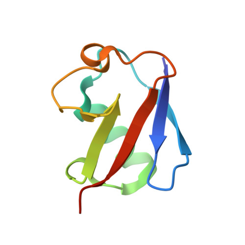
    The Papain-like protease (PLpro) is a domain of a multi-functional, non-structural protein 3 of coronaviruses. PLpro cleaves viral polyproteins and posttranslational conjugates with poly-ubiquitin and protective ISG15, composed of two ubiquitin-like (UBL) domains. Across coronaviruses, PLpro showed divergent selectivity for recognition and cleavage of posttranslational conjugates despite sequence conservation. We show that SARS-CoV-2 PLpro binds human ISG15 and K48-linked di-ubiquitin (K48-Ub 2 ) with nanomolar affinity and detect alternate weaker-binding modes. Crystal structures of untethered PLpro complexes with ISG15 and K48-Ub 2 combined with solution NMR and cross-linking mass spectrometry revealed how the two domains of ISG15 or K48-Ub 2 are differently utilized in interactions with PLpro. Analysis of protein interface energetics predicted differential binding stabilities of the two UBL/Ub domains that were validated experimentally. We emphasize how substrate recognition can be tuned to cleave specifically ISG15 or K48-Ub 2 modifications while retaining capacity to cleave mono-Ub conjugates. These results highlight alternative druggable surfaces that would inhibit PLpro function.


  • Organizational Affiliation
    • Molecular Biophysics Graduate Program, University of Texas Southwestern Medical Center, Dallas, TX, 75390, USA.

Macromolecule Content 

  • Total Structure Weight: 17.41 kDa 
  • Atom Count: 1,432 
  • Modeled Residue Count: 148 
  • Deposited Residue Count: 153 
  • Unique protein chains: 2

Macromolecules

Find similar proteins by:|  3D Structure
Entity ID: 1
MoleculeChains  Sequence LengthOrganismDetailsImage
Ubiquitin76Homo sapiensMutation(s): 1 
Gene Names: UBB
UniProt & NIH Common Fund Data Resources
Find proteins for P0CG47 (Homo sapiens)
Explore P0CG47 
Go to UniProtKB:  P0CG47
PHAROS:  P0CG47
GTEx:  ENSG00000170315 
Entity Groups
Sequence Clusters30% Identity50% Identity70% Identity90% Identity95% Identity100% Identity
UniProt GroupP0CG47
Sequence Annotations
Expand
Reference Sequence
Find similar proteins by:|  3D Structure
Entity ID: 2
MoleculeChains  Sequence LengthOrganismDetailsImage
Ubiquitin77Homo sapiensMutation(s): 0 
Gene Names: UBB
UniProt & NIH Common Fund Data Resources
Find proteins for P0CG47 (Homo sapiens)
Explore P0CG47 
Go to UniProtKB:  P0CG47
PHAROS:  P0CG47
GTEx:  ENSG00000170315 
Entity Groups
Sequence Clusters30% Identity50% Identity70% Identity90% Identity95% Identity100% Identity
UniProt GroupP0CG47
Sequence Annotations
Expand
Reference Sequence

Experimental Data & Validation

Experimental Data

  • Method: X-RAY DIFFRACTION
  • Resolution: 1.25 Å
  • R-Value Free:  0.175 (Depositor), 0.175 (DCC) 
  • R-Value Work:  0.129 (Depositor), 0.129 (DCC) 
  • R-Value Observed: 0.131 (Depositor) 
Space Group: P 1 21 1
Unit Cell:
Length ( Å )Angle ( ˚ )
a = 23.804α = 90
b = 56.577β = 93.37
c = 46.201γ = 90
Software Package:
Software NamePurpose
REFMACrefinement
HKL-3000data scaling
PDB_EXTRACTdata extraction
HKL-3000data reduction
HKL-3000phasing

Structure Validation

View Full Validation Report


Entry History 

& Funding Information

Deposition Data


Funding OrganizationLocationGrant Number
Department of Energy (DOE, United States)United StatesDE-AC02-06CH11357

Revision History  (Full details and data files)

  • Version 1.0: 2021-09-22
    Type: Initial release
  • Version 1.1: 2021-12-15
    Changes: Author supporting evidence, Structure summary
  • Version 2.0: 2022-03-16
    Type: Coordinate replacement
    Reason: Model orientation/position
    Changes: Advisory, Atomic model, Author supporting evidence, Data collection, Database references, Derived calculations, Non-polymer description, Polymer sequence, Refinement description, Source and taxonomy, Structure summary
  • Version 2.1: 2023-05-31
    Changes: Database references
  • Version 2.2: 2023-10-25
    Changes: Data collection, Refinement description