Structural basis of binding and inhibition of ornithine decarboxylase by 1-amino-oxy-3-aminopropane.
Zhou, X.E., Suino-Powell, K., Schultz, C.R., Aleiwi, B., Brunzelle, J.S., Lamp, J., Vega, I.E., Ellsworth, E., Bachmann, A.S., Melcher, K.(2021) Biochem J 478: 4137-4149
- PubMed: 34796899 Search on PubMedSearch on PubMed Central
- DOI: https://doi.org/10.1042/BCJ20210647
- Primary Citation Related Structures:
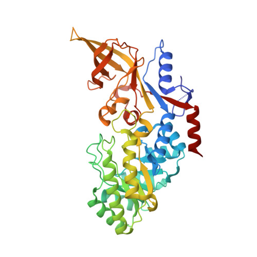
7S3F, 7S3G - PubMed Abstract:
Ornithine decarboxylase (ODC) is the rate-limiting enzyme for the synthesis of polyamines (PAs). PAs are oncometabolites that are required for proliferation, and pharmaceutical ODC inhibition is pursued for the treatment of hyperproliferative diseases, including cancer and infectious diseases. The most potent ODC inhibitor is 1-amino-oxy-3-aminopropane (APA). A previous crystal structure of an ODC-APA complex indicated that APA non-covalently binds ODC and its cofactor pyridoxal 5-phosphate (PLP) and functions by competing with the ODC substrate ornithine for binding to the catalytic site. We have revisited the mechanism of APA binding and ODC inhibition through a new crystal structure of APA-bound ODC, which we solved at 2.49 Å resolution. The structure unambiguously shows the presence of a covalent oxime between APA and PLP in the catalytic site, which we confirmed in solution by mass spectrometry. The stable oxime makes extensive interactions with ODC but cannot be catabolized, explaining APA's high potency in ODC inhibition. In addition, we solved an ODC/PLP complex structure with citrate bound at the substrate-binding pocket. These two structures provide new structural scaffolds for developing more efficient pharmaceutical ODC inhibitors.
- Department of Structural Biology, Van Andel Institute, Grand Rapids, Michigan 49503, U.S.A.
Organizational Affiliation: