7RX0

Complex of AMPPNP-Kif7 and Gli2 Zinc-Finger domain bound to microtubules


Experimental Data Snapshot

  • Method: ELECTRON MICROSCOPY
  • Resolution: 3.89 Å
  • Aggregation State: HELICAL ARRAY 
  • Reconstruction Method: HELICAL 

wwPDB Validation   3D Report Full Report


This is version 1.3 of the entry. See complete history


Literature

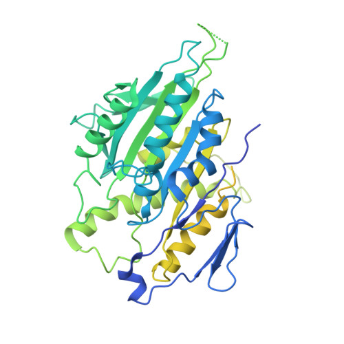
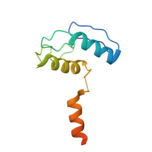
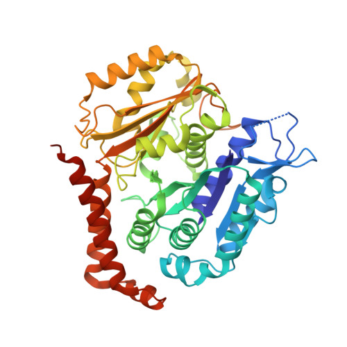
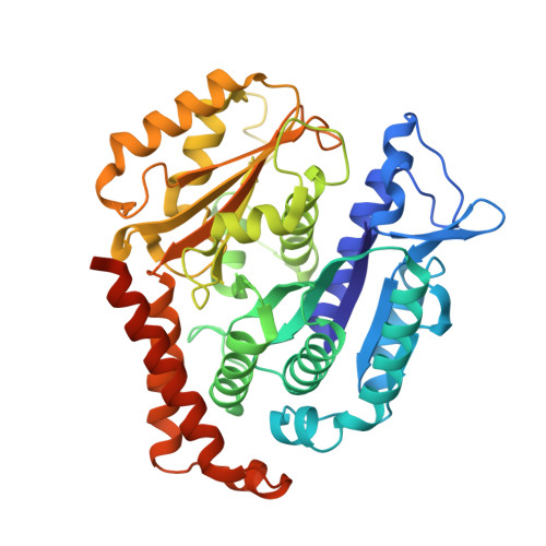
Cytoskeletal regulation of a transcription factor by DNA mimicry via coiled-coil interactions.

Haque, F.Freniere, C.Ye, Q.Mani, N.Wilson-Kubalek, E.M.Ku, P.I.Milligan, R.A.Subramanian, R.

(2022) Nat Cell Biol 24: 1088-1098

  • DOI: https://doi.org/10.1038/s41556-022-00935-7
  • Primary Citation of Related Structures:  
    7RX0

  • PubMed Abstract: 

    A long-established strategy for transcription regulation is the tethering of transcription factors to cellular membranes. By contrast, the principal effectors of Hedgehog signalling, the GLI transcription factors, are regulated by microtubules in the primary cilium and the cytoplasm. How GLI is tethered to microtubules remains unclear. Here, we uncover DNA mimicry by the ciliary kinesin KIF7 as a mechanism for the recruitment of GLI to microtubules, wherein the coiled-coil dimerization domain of KIF7, characterized by its striking shape, size and charge similarity to DNA, forms a complex with the DNA-binding zinc fingers in GLI, thus revealing a mode of tethering a DNA-binding protein to the cytoskeleton. GLI increases KIF7 microtubule affinity and consequently modulates the localization of both proteins to microtubules and the cilium tip. Thus, the kinesin-microtubule system is not a passive GLI tether but a regulatable platform tuned by the kinesin-transcription factor interaction. We retooled this coiled-coil-based GLI-KIF7 interaction to inhibit the nuclear and cilium localization of GLI. This strategy can potentially be exploited to downregulate erroneously activated GLI in human cancers.


  • Organizational Affiliation

    Department of Molecular Biology, Massachusetts General Hospital, Boston, MA, USA.


Macromolecules
Find similar proteins by:  (by identity cutoff)  |  3D Structure
Entity ID: 1
MoleculeChains Sequence LengthOrganismDetailsImage
Kinesin-like protein KIF7A [auth C]544Homo sapiensMutation(s): 0 
Gene Names: KIF7UNQ340/PRO539
UniProt & NIH Common Fund Data Resources
Find proteins for Q2M1P5 (Homo sapiens)
Explore Q2M1P5 
Go to UniProtKB:  Q2M1P5
PHAROS:  Q2M1P5
GTEx:  ENSG00000166813 
Entity Groups  
Sequence Clusters30% Identity50% Identity70% Identity90% Identity95% Identity100% Identity
UniProt GroupQ2M1P5
Sequence Annotations
Expand
  • Reference Sequence
Find similar proteins by:  (by identity cutoff)  |  3D Structure
Entity ID: 2
MoleculeChains Sequence LengthOrganismDetailsImage
Zinc finger protein GLI2B [auth G]92Homo sapiensMutation(s): 0 
Sequence Annotations
Expand
  • Reference Sequence
Find similar proteins by:  (by identity cutoff)  |  3D Structure
Entity ID: 3
MoleculeChains Sequence LengthOrganismDetailsImage
Tubulin alpha-1A chainC [auth A]451Sus scrofaMutation(s): 0 
UniProt
Find proteins for P02550 (Sus scrofa)
Explore P02550 
Go to UniProtKB:  P02550
Entity Groups  
Sequence Clusters30% Identity50% Identity70% Identity90% Identity95% Identity100% Identity
UniProt GroupP02550
Sequence Annotations
Expand
  • Reference Sequence
Find similar proteins by:  (by identity cutoff)  |  3D Structure
Entity ID: 4
MoleculeChains Sequence LengthOrganismDetailsImage
Tubulin beta chainD [auth B]445Sus scrofaMutation(s): 0 
UniProt
Find proteins for P02554 (Sus scrofa)
Explore P02554 
Go to UniProtKB:  P02554
Entity Groups  
Sequence Clusters30% Identity50% Identity70% Identity90% Identity95% Identity100% Identity
UniProt GroupP02554
Sequence Annotations
Expand
  • Reference Sequence
Small Molecules
Ligands 4 Unique
IDChains Name / Formula / InChI Key2D Diagram3D Interactions
GTP
Query on GTP

Download Ideal Coordinates CCD File 
F [auth A]GUANOSINE-5'-TRIPHOSPHATE
C10 H16 N5 O14 P3
XKMLYUALXHKNFT-UUOKFMHZSA-N
G2P
Query on G2P

Download Ideal Coordinates CCD File 
G [auth B]PHOSPHOMETHYLPHOSPHONIC ACID GUANYLATE ESTER
C11 H18 N5 O13 P3
GXTIEXDFEKYVGY-KQYNXXCUSA-N
ANP
Query on ANP

Download Ideal Coordinates CCD File 
E [auth C]PHOSPHOAMINOPHOSPHONIC ACID-ADENYLATE ESTER
C10 H17 N6 O12 P3
PVKSNHVPLWYQGJ-KQYNXXCUSA-N
MG
Query on MG

Download Ideal Coordinates CCD File 
H [auth B]MAGNESIUM ION
Mg
JLVVSXFLKOJNIY-UHFFFAOYSA-N
Experimental Data & Validation

Experimental Data

  • Method: ELECTRON MICROSCOPY
  • Resolution: 3.89 Å
  • Aggregation State: HELICAL ARRAY 
  • Reconstruction Method: HELICAL 
EM Software:
TaskSoftware PackageVersion
RECONSTRUCTIONFREALIX
MODEL REFINEMENTPHENIX1.17.1-3660

Structure Validation

View Full Validation Report


Entry History & Funding Information

Deposition Data


Funding OrganizationLocationGrant Number
American Cancer SocietyUnited States--

Revision History  (Full details and data files)

  • Version 1.0: 2022-06-15
    Type: Initial release
  • Version 1.1: 2022-06-29
    Changes: Database references
  • Version 1.2: 2022-07-06
    Changes: Database references
  • Version 1.3: 2022-07-27
    Changes: Database references