


Bromodomain-containing protein 4 (BRD4) bromodomain 2 (BD2) complexed with XR844
Xiong, R., Ratia, K.M., Li, Y., Shen, Z., Zhao, J., Huang, F., Dubrovyskyii, O., Thatcher, G.R.To be published.
Experimental Data Snapshot
Starting Model: experimental
View more details
Entity ID: 1 | |||||
|---|---|---|---|---|---|
| Molecule | Chains | Sequence Length | Organism | Details | Image |
| Bromodomain-containing protein 4 | 130 | Homo sapiens | Mutation(s): 0 Gene Names: BRD4, HUNK1 | 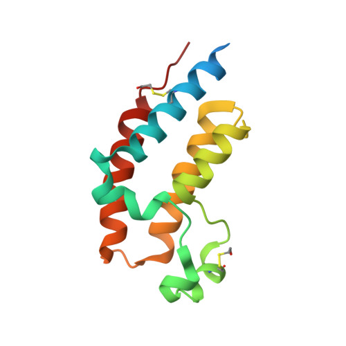 | |
UniProt & NIH Common Fund Data Resources | |||||
PHAROS: O60885 GTEx: ENSG00000141867 | |||||
Entity Groups | |||||
| Sequence Clusters | 30% Identity50% Identity70% Identity90% Identity95% Identity100% Identity | ||||
| UniProt Group | O60885 | ||||
Sequence AnnotationsExpand | |||||
Reference Sequence | |||||
| Ligands 1 Unique | |||||
|---|---|---|---|---|---|
| ID | Chains | Name / Formula / InChI Key | 2D Diagram | 3D Interactions | |
| 7QZ (Subject of Investigation/LOI) Download:Ideal Coordinates CCD File | B [auth A] | N-{1-[1,1-di(pyridin-2-yl)ethyl]-6-(5-{[(2-fluorophenyl)carbamoyl]amino}-1-methyl-6-oxo-1,6-dihydropyridin-3-yl)-1H-indol-4-yl}-2,2,2-trifluoroethane-1-sulfonamide C35 H29 F4 N7 O4 S RFUGSFUWHIKRES-UHFFFAOYSA-N |  | ||
| Modified Residues 1 Unique | |||||
|---|---|---|---|---|---|
| ID | Chains | Type | Formula | 2D Diagram | Parent |
| CME Query on CME | A | L-PEPTIDE LINKING | C5 H11 N O3 S2 |  | CYS |
| Length ( Å ) | Angle ( ˚ ) |
|---|---|
| a = 32.98 | α = 90 |
| b = 53.168 | β = 90 |
| c = 72.481 | γ = 90 |
| Software Name | Purpose |
|---|---|
| XDS | data reduction |
| Aimless | data scaling |
| REFMAC | refinement |
| PDB_EXTRACT | data extraction |
| MOLREP | phasing |
| Funding Organization | Location | Grant Number |
|---|---|---|
| National Institutes of Health/National Center for Research Resources (NIH/NCRR) | United States | UL1RR029879 |


