7RQZ

Cryo-EM structure of the full-length TRPV1 with RTx at 48 degrees Celsius, in an open state, class alpha


Experimental Data Snapshot

  • Method: ELECTRON MICROSCOPY
  • Resolution: 3.32 Å
  • Aggregation State: PARTICLE 
  • Reconstruction Method: SINGLE PARTICLE 

wwPDB Validation   3D Report Full Report


This is version 1.0 of the entry. See complete history


Literature

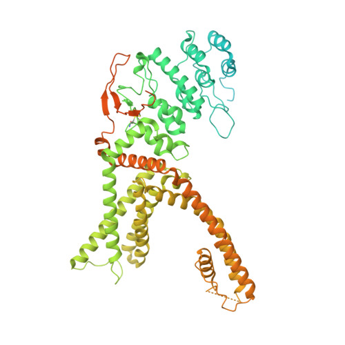
Vanilloid-dependent TRPV1 opening trajectory from cryoEM ensemble analysis.

Kwon, D.H.Zhang, F.Fedor, J.G.Suo, Y.Lee, S.Y.

(2022) Nat Commun 13: 2874-2874

  • DOI: https://doi.org/10.1038/s41467-022-30602-2
  • Primary Citation of Related Structures:  
    7RQU, 7RQV, 7RQW, 7RQX, 7RQY, 7RQZ

  • PubMed Abstract: 

    Single particle cryo-EM often yields multiple protein conformations within a single dataset, but experimentally deducing the temporal relationship of these conformers within a conformational trajectory is not trivial. Here, we use thermal titration methods and cryo-EM in an attempt to obtain temporal resolution of the conformational trajectory of the vanilloid receptor TRPV1 with resiniferatoxin (RTx) bound. Based on our cryo-EM ensemble analysis, RTx binding to TRPV1 appears to induce intracellular gate opening first, followed by selectivity filter dilation, then pore loop rearrangement to reach the final open state. This apparent conformational wave likely arises from the concerted, stepwise, additive structural changes of TRPV1 over many subdomains. Greater understanding of the RTx-mediated long-range allostery of TRPV1 could help further the therapeutic potential of RTx, which is a promising drug candidate for pain relief associated with advanced cancer or knee arthritis.


  • Organizational Affiliation

    Department of Biochemistry, Duke University School of Medicine, Durham, NC, 27710, USA.


Macromolecules
Find similar proteins by:  (by identity cutoff)  |  3D Structure
Entity ID: 1
MoleculeChains Sequence LengthOrganismDetailsImage
Transient receptor potential cation channel subfamily V member 1A,
B [auth C],
C [auth B],
D
868Rattus norvegicusMutation(s): 0 
Gene Names: Trpv1Vr1Vr1l
Membrane Entity: Yes 
UniProt
Find proteins for O35433 (Rattus norvegicus)
Explore O35433 
Go to UniProtKB:  O35433
Entity Groups  
Sequence Clusters30% Identity50% Identity70% Identity90% Identity95% Identity100% Identity
UniProt GroupO35433
Sequence Annotations
Expand
  • Reference Sequence
Small Molecules
Ligands 3 Unique
IDChains Name / Formula / InChI Key2D Diagram3D Interactions
LBN
Query on LBN

Download Ideal Coordinates CCD File 
F [auth A]
I [auth A]
K [auth C]
N [auth C]
P [auth B]
F [auth A],
I [auth A],
K [auth C],
N [auth C],
P [auth B],
S [auth B],
T [auth D],
V [auth D]
1-palmitoyl-2-oleoyl-sn-glycero-3-phosphocholine
C42 H82 N O8 P
WTJKGGKOPKCXLL-VYOBOKEXSA-N
6OU
Query on 6OU

Download Ideal Coordinates CCD File 
G [auth A]
H [auth A]
L [auth C]
M [auth C]
Q [auth B]
G [auth A],
H [auth A],
L [auth C],
M [auth C],
Q [auth B],
R [auth B],
W [auth D],
X [auth D]
[(2~{R})-1-[2-azanylethoxy(oxidanyl)phosphoryl]oxy-3-hexadecanoyloxy-propan-2-yl] (~{Z})-octadec-9-enoate
C39 H76 N O8 P
FHQVHHIBKUMWTI-OTMQOFQLSA-N
6EU
Query on 6EU

Download Ideal Coordinates CCD File 
E [auth A],
J [auth C],
O [auth B],
U [auth D]
resiniferatoxin
C37 H40 O9
DSDNAKHZNJAGHN-MXTYGGKSSA-N
Experimental Data & Validation

Experimental Data

  • Method: ELECTRON MICROSCOPY
  • Resolution: 3.32 Å
  • Aggregation State: PARTICLE 
  • Reconstruction Method: SINGLE PARTICLE 
EM Software:
TaskSoftware PackageVersion
MODEL REFINEMENTPHENIX1.18.2
RECONSTRUCTIONRELION

Structure Validation

View Full Validation Report


Entry History & Funding Information

Deposition Data


Funding OrganizationLocationGrant Number
National Institutes of Health/National Institute of Neurological Disorders and Stroke (NIH/NINDS)United StatesR35NS097241

Revision History  (Full details and data files)

  • Version 1.0: 2022-06-01
    Type: Initial release