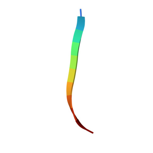
7ROJ

Amyloid-related segment of alphaB-crystallin residues 90-100 with G95W mutation


Experimental Data Snapshot

  • Method: X-RAY DIFFRACTION
  • Resolution: 1.60 Å
  • R-Value Free: 0.277 
  • R-Value Work: 0.239 
  • R-Value Observed: 0.243 

wwPDB Validation   3D Report Full Report


This is version 1.2 of the entry. See complete history


Literature

Atomic view of an amyloid dodecamer exhibiting selective cellular toxic vulnerability in acute brain slices.

Gray, A.L.H.Sawaya, M.R.Acharyya, D.Lou, J.Edington, E.M.Best, M.D.Prosser, R.A.Eisenberg, D.S.Do, T.D.

(2022) Protein Sci 31: 716-727

  • DOI: https://doi.org/10.1002/pro.4268
  • Primary Citation of Related Structures:  
    7ROJ, 7ROL

  • PubMed Abstract: 

    Atomic structures of amyloid oligomers that capture the neurodegenerative disease pathology are essential to understand disease-state causes and finding cures. Here we investigate the G6W mutation of the cytotoxic, hexameric amyloid model KV11. The mutation results into an asymmetric dodecamer composed of a pair of 30° twisted antiparallel β-sheets. The complete break between adjacent β-strands is unprecedented among amyloid fibril crystal structures and supports that our structure is an oligomer. The poor shape complementarity between mated sheets reveals an interior channel for binding lipids, suggesting that the toxicity may be due to a perturbation of lipid transport rather than a direct disruption of membrane integrity. Viability assays on mouse suprachiasmatic nucleus, anterior hypothalamus, and cerebral cortex demonstrated selective regional vulnerability consistent with Alzheimer's disease. Neuropeptides released from the brain slices may provide clues to how G6W initiates cellular injury.


  • Organizational Affiliation

    Department of Chemistry, University of Tennessee, Knoxville, Tennessee, USA.


Macromolecules

Find similar proteins by:  Sequence   |   3D Structure  

Entity ID: 1
MoleculeChains Sequence LengthOrganismDetailsImage
Alpha-crystallin B chain peptide
A, B, C, D, E
A, B, C, D, E, F, G, H, I, J, K, L
11Homo sapiensMutation(s): 1 
UniProt & NIH Common Fund Data Resources
Find proteins for P02511 (Homo sapiens)
Explore P02511 
Go to UniProtKB:  P02511
PHAROS:  P02511
GTEx:  ENSG00000109846 
Entity Groups  
Sequence Clusters30% Identity50% Identity70% Identity90% Identity95% Identity100% Identity
UniProt GroupP02511
Sequence Annotations
Expand
  • Reference Sequence
Experimental Data & Validation

Experimental Data

  • Method: X-RAY DIFFRACTION
  • Resolution: 1.60 Å
  • R-Value Free: 0.277 
  • R-Value Work: 0.239 
  • R-Value Observed: 0.243 
  • Space Group: P 1 21 1
Unit Cell:
Length ( Å )Angle ( ˚ )
a = 25.27α = 90
b = 36.33β = 91.92
c = 64.13γ = 90
Software Package:
Software NamePurpose
PHENIXrefinement
XDSdata reduction
XSCALEdata scaling
PHASERphasing
PDB_EXTRACTdata extraction

Structure Validation

View Full Validation Report


Entry History & Funding Information

Deposition Data


Funding OrganizationLocationGrant Number
Howard Hughes Medical Institute (HHMI)United States--

Revision History  (Full details and data files)

  • Version 1.0: 2022-01-12
    Type: Initial release
  • Version 1.1: 2022-03-02
    Changes: Database references
  • Version 1.2: 2023-10-18
    Changes: Data collection, Refinement description