7REJ | pdb_00007rej

Tailspike protein 4 (TSP4) from phage CBA120, residues 1-335, obtained in the presence of NaK-Tartrate


Experimental Data Snapshot

  • Method: X-RAY DIFFRACTION
  • Resolution: 2.60 Å
  • R-Value Free: 
    0.229 (Depositor), 0.229 (DCC) 
  • R-Value Work: 
    0.204 (Depositor), 0.206 (DCC) 
  • R-Value Observed: 
    0.206 (Depositor) 

Starting Model: experimental
View more details

wwPDB Validation 3D Report Full Report

Validation slider image for 7REJ

This is version 1.1 of the entry. See complete history

Literature

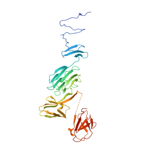
Structure of Escherichia coli O157:H7 bacteriophage CBA120 tailspike protein 4 baseplate anchor and tailspike assembly domains (TSP4-N).

Chao, K.L.Shang, X.Greenfield, J.Linden, S.B.Alreja, A.B.Nelson, D.C.Herzberg, O.

(2022) Sci Rep 12: 2061-2061

  • DOI: https://doi.org/10.1038/s41598-022-06073-2
  • Primary Citation Related Structures: 
    7REJ, 7RFO, 7RFV

  • PubMed Abstract: 

    Four tailspike proteins (TSP1-4) of Escherichia coli O157:H7 bacteriophage CBA120 enable infection of multiple hosts. They form a branched complex that attaches to the tail baseplate. Each TSP recognizes a different lipopolysaccharide on the membrane of a different bacterial host. The 335 N-terminal residues of TSP4 promote the assembly of the TSP complex and anchor it to the tail baseplate. The crystal structure of TSP4-N 335 reveals a trimeric protein comprising four domains. The baseplate anchor domain (AD) contains an intertwined triple-stranded β-helix. The ensuing XD1, XD2 and XD3 β-sheet containing domains mediate the binding of TSP1-3 to TSP4. Each of the XD domains adopts the same fold as the respective XD domains of bacteriophage T4 gp10 baseplate protein, known to engage in protein-protein interactions via its XD2 and XD3 domains. The structural similarity suggests that XD2 and XD3 of TSP4 also function in protein-protein interactions. Analytical ultracentrifugation analyses of TSP4-N 335 and of domain deletion proteins showed how TSP4-N 335 promotes the formation of the TSP quaternary complex. TSP1 and TSP2 bind directly to TSP4 whereas TSP3 binding requires a pre-formed TSP4-N 335 :TSP2 complex. A 3-dimensional model of the bacteriophage CBA120 TSP complex has been developed based on the structural and ultracentrifuge information.


  • Organizational Affiliation
    • Institute for Bioscience and Biotechnology Research, University of Maryland, 9600 Gudelsky Drive, Rockville, MD, 20850, USA.

Macromolecule Content 

  • Total Structure Weight: 72.78 kDa 
  • Atom Count: 4,322 
  • Modeled Residue Count: 576 
  • Deposited Residue Count: 682 
  • Unique protein chains: 1

Macromolecules

Find similar proteins by:|  3D Structure
Entity ID: 1
MoleculeChains  Sequence LengthOrganismDetailsImage
Tailspike protein
A, B
341Kuttervirus CBA120Mutation(s): 0 
Gene Names: orf213
UniProt
Find proteins for G3M192 (Escherichia phage Cba120)
Explore G3M192 
Go to UniProtKB:  G3M192
Entity Groups
Sequence Clusters30% Identity50% Identity70% Identity90% Identity95% Identity100% Identity
UniProt GroupG3M192
Sequence Annotations
Expand
Reference Sequence

Experimental Data & Validation

Experimental Data

  • Method: X-RAY DIFFRACTION
  • Resolution: 2.60 Å
  • R-Value Free:  0.229 (Depositor), 0.229 (DCC) 
  • R-Value Work:  0.204 (Depositor), 0.206 (DCC) 
  • R-Value Observed: 0.206 (Depositor) 
Space Group: H 3
Unit Cell:
Length ( Å )Angle ( ˚ )
a = 78.03α = 90
b = 78.03β = 90
c = 326.87γ = 120
Software Package:
Software NamePurpose
REFMACrefinement
PDB_EXTRACTdata extraction
XDSdata reduction
XDSdata scaling
PHASERphasing

Structure Validation

View Full Validation Report


Entry History 

& Funding Information

Deposition Data


Funding OrganizationLocationGrant Number
National Institutes of Health/National Institute of General Medical Sciences (NIH/NIGMS)United StatesRO1GM110202

Revision History  (Full details and data files)

  • Version 1.0: 2022-02-23
    Type: Initial release
  • Version 1.1: 2024-04-03
    Changes: Data collection, Refinement description