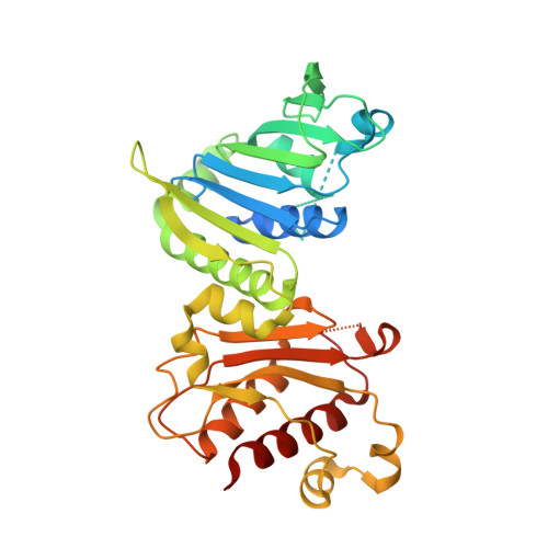
PMS2 variant results in loss of ATPase activity without compromising mismatch repair.
D'Arcy, B.M., Arrington, J., Weisman, J., McClellan, S.B., Yang, Z., Deivanayagam, C., Blount, J., Prakash, A.(2022) Mol Genet Genomic Med 10: e1908-e1908
- PubMed: 35189042 Search on PubMedSearch on PubMed Central
- DOI: https://doi.org/10.1002/mgg3.1908
- Primary Citation Related Structures:
7RCB, 7RCI, 7RCK - PubMed Abstract:
Hereditary cancer syndromes account for approximately 5%-10% of all diagnosed cancer cases. Lynch syndrome (LS) is an autosomal dominant hereditary cancer condition that predisposes individuals to an elevated lifetime risk for developing colorectal, endometrial, and other cancers. LS results from a pathogenic mutation in one of four mismatch repair (MMR) genes (MSH2, MSH6, MLH1, and PMS2). The diagnosis of LS is often challenged by the identification of missense mutations, termed variants of uncertain significance, whose functional effect on the protein is not known. Of the eight PMS2 variants initially selected for this study, we identified a variant within the N-terminal domain where asparagine 335 is mutated to serine, p.Asn335Ser, which lacked ATPase activity, yet appears to be proficient in MMR. To expand our understanding of this functional dichotomy, we performed biophysical and structural studies, and noted that p.Asn335Ser binds to ATP but is unable to hydrolyze it to ADP. To examine the impact of p.Asn335Ser on MMR, we developed a novel in-cell fluorescent-based microsatellite instability reporter that revealed p.Asn335Ser maintained genomic stability. We conclude that in the absence of gross structural changes, PMS2 ATP hydrolysis is not necessary for proficient MMR and that the ATPase deficient p.Asn335Ser variant is likely benign.
- Mitchell Cancer Institute, University of South Alabama Health, Mobile, Alabama, USA.
Organizational Affiliation: