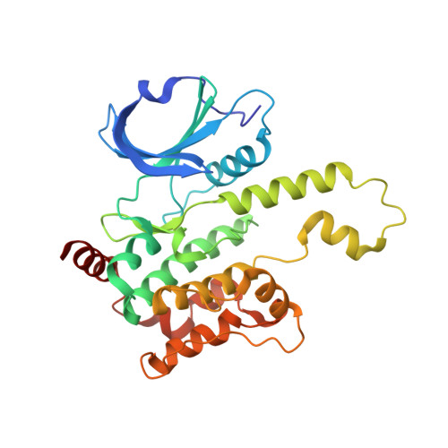
7R9N

Crystal structure of HPK1 in complex with GNE1858


Experimental Data Snapshot

  • Method: X-RAY DIFFRACTION
  • Resolution: 1.50 Å
  • R-Value Free: 0.191 
  • R-Value Work: 0.166 
  • R-Value Observed: 0.167 

wwPDB Validation   3D Report Full Report


Ligand Structure Quality Assessment 


This is version 1.2 of the entry. See complete history


Literature

Discovery of Spiro-azaindoline Inhibitors of Hematopoietic Progenitor Kinase 1 (HPK1).

Chan, B.K.Seward, E.Lainchbury, M.Brewer, T.F.An, L.Blench, T.Cartwright, M.W.Chan, G.K.Y.Choo, E.F.Drummond, J.Elliott, R.L.Gancia, E.Gazzard, L.Hu, B.Jones, G.E.Luo, X.Madin, A.Malhotra, S.Moffat, J.G.Pang, J.Salphati, L.Sneeringer, C.J.Stivala, C.E.Wei, B.Wang, W.Wu, P.Heffron, T.P.

(2022) ACS Med Chem Lett 13: 84-91

  • DOI: https://doi.org/10.1021/acsmedchemlett.1c00473
  • Primary Citation of Related Structures:  
    7R9L, 7R9N, 7R9P, 7R9T

  • PubMed Abstract: 

    Hematopoietic progenitor kinase 1 (HPK1) is implicated as a negative regulator of T-cell receptor-induced T-cell activation. Studies using HPK1 kinase-dead knock-in animals have demonstrated the loss of HPK1 kinase activity resulted in an increase in T-cell function and tumor growth inhibition in glioma models. Herein, we describe the discovery of a series of small molecule inhibitors of HPK1. Using a structure-based drug design approach, the kinase selectivity of the molecules was significantly improved by inducing and stabilizing an unusual P-loop folded binding mode. The metabolic liabilities of the initial 7-azaindole high-throughput screening hit were mitigated by addressing a key metabolic soft spot along with physicochemical property-based optimization. The resulting spiro-azaindoline HPK1 inhibitors demonstrated improved in vitro ADME properties and the ability to induce cytokine production in primary human T-cells.


  • Organizational Affiliation

    Genentech Inc., 1 DNA Way, South San Francisco, California 94080, United States.


Macromolecules
Find similar proteins by:  (by identity cutoff)  |  3D Structure
Entity ID: 1
MoleculeChains Sequence LengthOrganismDetailsImage
Hematopoietic progenitor kinase
A, B
297Homo sapiensMutation(s): 0 
Gene Names: MAP4K1HPK1
EC: 2.7.11.1
UniProt & NIH Common Fund Data Resources
Find proteins for Q92918 (Homo sapiens)
Explore Q92918 
Go to UniProtKB:  Q92918
PHAROS:  Q92918
GTEx:  ENSG00000104814 
Entity Groups  
Sequence Clusters30% Identity50% Identity70% Identity90% Identity95% Identity100% Identity
UniProt GroupQ92918
Sequence Annotations
Expand
  • Reference Sequence
Small Molecules
Ligands 3 Unique
IDChains Name / Formula / InChI Key2D Diagram3D Interactions
F97 (Subject of Investigation/LOI)
Query on F97

Download Ideal Coordinates CCD File 
C [auth A],
E [auth B]
N-{2-(3,3-difluoropyrrolidin-1-yl)-6-[(3R)-pyrrolidin-3-yl]pyrimidin-4-yl}-1-(propan-2-yl)-1H-pyrazolo[4,3-c]pyridin-6-amine
C21 H26 F2 N8
JKPAJCPZQIJPPS-CQSZACIVSA-N
MES
Query on MES

Download Ideal Coordinates CCD File 
F [auth B]2-(N-MORPHOLINO)-ETHANESULFONIC ACID
C6 H13 N O4 S
SXGZJKUKBWWHRA-UHFFFAOYSA-N
EDO
Query on EDO

Download Ideal Coordinates CCD File 
D [auth A]1,2-ETHANEDIOL
C2 H6 O2
LYCAIKOWRPUZTN-UHFFFAOYSA-N
Experimental Data & Validation

Experimental Data

  • Method: X-RAY DIFFRACTION
  • Resolution: 1.50 Å
  • R-Value Free: 0.191 
  • R-Value Work: 0.166 
  • R-Value Observed: 0.167 
  • Space Group: P 1
Unit Cell:
Length ( Å )Angle ( ˚ )
a = 52.006α = 70.36
b = 57.963β = 66.43
c = 61.562γ = 68.57
Software Package:
Software NamePurpose
Aimlessdata scaling
PHENIXrefinement
PDB_EXTRACTdata extraction
XDSdata reduction
PHASERphasing

Structure Validation

View Full Validation Report


Ligand Structure Quality Assessment 


Entry History 

Deposition Data

Revision History  (Full details and data files)

  • Version 1.0: 2022-01-05
    Type: Initial release
  • Version 1.1: 2022-02-09
    Changes: Database references
  • Version 1.2: 2023-10-18
    Changes: Data collection, Refinement description