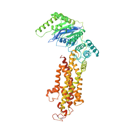
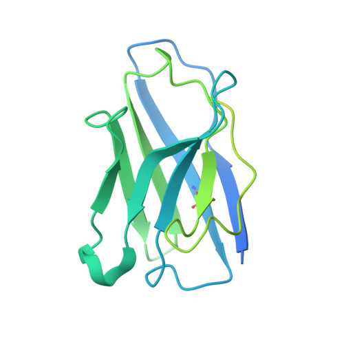
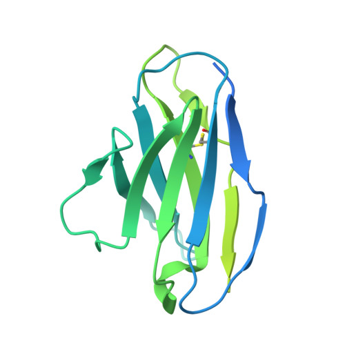
7R8A

The structure of human ABCG5/ABCG8 purified from mammalian cells


Experimental Data Snapshot

  • Method: ELECTRON MICROSCOPY
  • Resolution: 2.90 Å
  • Aggregation State: PARTICLE 
  • Reconstruction Method: SINGLE PARTICLE 

wwPDB Validation   3D Report Full Report


This is version 1.0 of the entry. See complete history


Literature

Molecular basis of cholesterol efflux via ABCG subfamily transporters.

Sun, Y.Wang, J.Long, T.Qi, X.Donnelly, L.Elghobashi-Meinhardt, N.Esparza, L.Cohen, J.C.Xie, X.S.Hobbs, H.H.Li, X.

(2021) Proc Natl Acad Sci U S A 118

  • DOI: https://doi.org/10.1073/pnas.2110483118
  • Primary Citation of Related Structures:  
    7R87, 7R88, 7R89, 7R8A, 7R8B, 7R8C, 7R8D, 7R8E

  • PubMed Abstract: 

    The ABCG1 homodimer (G1) and ABCG5-ABCG8 heterodimer (G5G8), two members of the adenosine triphosphate (ATP)-binding cassette (ABC) transporter G family, are required for maintenance of cellular cholesterol levels. G5G8 mediates secretion of neutral sterols into bile and the gut lumen, whereas G1 transports cholesterol from macrophages to high-density lipoproteins (HDLs). The mechanisms used by G5G8 and G1 to recognize and export sterols remain unclear. Here, we report cryoelectron microscopy (cryo-EM) structures of human G5G8 in sterol-bound and human G1 in cholesterol- and ATP-bound states. Both transporters have a sterol-binding site that is accessible from the cytosolic leaflet. A second site is present midway through the transmembrane domains of G5G8. The Walker A motif of G8 adopts a unique conformation that accounts for the marked asymmetry in ATPase activities between the two nucleotide-binding sites of G5G8. These structures, along with functional validation studies, provide a mechanistic framework for understanding cholesterol efflux via ABC transporters.


  • Organizational Affiliation

    Department of Molecular Genetics, University of Texas Southwestern Medical Center, Dallas, TX 75390.


Macromolecules
Find similar proteins by:  (by identity cutoff)  |  3D Structure
Entity ID: 1
MoleculeChains Sequence LengthOrganismDetailsImage
ATP-binding cassette sub-family G member 5666Homo sapiensMutation(s): 0 
Gene Names: ABCG5
EC: 7.6.2
Membrane Entity: Yes 
UniProt & NIH Common Fund Data Resources
Find proteins for Q9H222 (Homo sapiens)
Explore Q9H222 
Go to UniProtKB:  Q9H222
PHAROS:  Q9H222
GTEx:  ENSG00000138075 
Entity Groups  
Sequence Clusters30% Identity50% Identity70% Identity90% Identity95% Identity100% Identity
UniProt GroupQ9H222
Sequence Annotations
Expand
  • Reference Sequence
Find similar proteins by:  (by identity cutoff)  |  3D Structure
Entity ID: 2
MoleculeChains Sequence LengthOrganismDetailsImage
ATP-binding cassette sub-family G member 8715Homo sapiensMutation(s): 0 
Gene Names: ABCG8
EC: 7.6.2
Membrane Entity: Yes 
UniProt & NIH Common Fund Data Resources
Find proteins for Q9H221 (Homo sapiens)
Explore Q9H221 
Go to UniProtKB:  Q9H221
PHAROS:  Q9H221
GTEx:  ENSG00000143921 
Entity Groups  
Sequence Clusters30% Identity50% Identity70% Identity90% Identity95% Identity100% Identity
UniProt GroupQ9H221
Sequence Annotations
Expand
  • Reference Sequence
Find similar proteins by:  (by identity cutoff)  |  3D Structure
Entity ID: 3
MoleculeChains Sequence LengthOrganismDetailsImage
2C7 Fab heavy chain245Mus musculusMutation(s): 0 
Entity Groups  
Sequence Clusters30% Identity50% Identity70% Identity90% Identity95% Identity100% Identity
Sequence Annotations
Expand
  • Reference Sequence
Find similar proteins by:  (by identity cutoff)  |  3D Structure
Entity ID: 4
MoleculeChains Sequence LengthOrganismDetailsImage
2C7 Fab light chain234Mus musculusMutation(s): 0 
Entity Groups  
Sequence Clusters30% Identity50% Identity70% Identity90% Identity95% Identity100% Identity
Sequence Annotations
Expand
  • Reference Sequence
Small Molecules
Ligands 1 Unique
IDChains Name / Formula / InChI Key2D Diagram3D Interactions
CLR (Subject of Investigation/LOI)
Query on CLR

Download Ideal Coordinates CCD File 
E [auth A]CHOLESTEROL
C27 H46 O
HVYWMOMLDIMFJA-DPAQBDIFSA-N
Experimental Data & Validation

Experimental Data

  • Method: ELECTRON MICROSCOPY
  • Resolution: 2.90 Å
  • Aggregation State: PARTICLE 
  • Reconstruction Method: SINGLE PARTICLE 

Structure Validation

View Full Validation Report


Entry History & Funding Information

Deposition Data


Funding OrganizationLocationGrant Number
National Institutes of Health/National Institute of General Medical Sciences (NIH/NIGMS)United States--
Welch FoundationUnited States--
Damon Runyon Cancer Research FoundationUnited States--

Revision History  (Full details and data files)

  • Version 1.0: 2021-09-08
    Type: Initial release