7R7T

p47-bound p97-R155H mutant with ADP


Experimental Data Snapshot

  • Method: ELECTRON MICROSCOPY
  • Resolution: 4.50 Å
  • Aggregation State: PARTICLE 
  • Reconstruction Method: SINGLE PARTICLE 

wwPDB Validation   3D Report Full Report


This is version 1.1 of the entry. See complete history


Literature

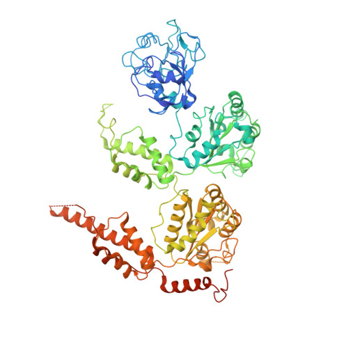
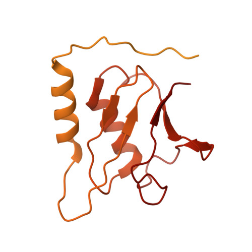
Structural and Functional Analysis of Disease-Linked p97 ATPase Mutant Complexes.

Nandi, P.Li, S.Columbres, R.C.A.Wang, F.Williams, D.R.Poh, Y.P.Chou, T.F.Chiu, P.L.

(2021) Int J Mol Sci 22

  • DOI: https://doi.org/10.3390/ijms22158079
  • Primary Citation of Related Structures:  
    7L5W, 7L5X, 7R7S, 7R7T, 7R7U

  • PubMed Abstract: 

    IBMPFD/ALS is a genetic disorder caused by a single amino acid mutation on the p97 ATPase, promoting ATPase activity and cofactor dysregulation. The disease mechanism underlying p97 ATPase malfunction remains unclear. To understand how the mutation alters the ATPase regulation, we assembled a full-length p97 R155H with its p47 cofactor and first visualized their structures using single-particle cryo-EM. More than one-third of the population was the dodecameric form. Nucleotide presence dissociates the dodecamer into two hexamers for its highly elevated function. The N-domains of the p97 R155H mutant all show up configurations in ADP- or ATP γ S-bound states. Our functional and structural analyses showed that the p47 binding is likely to impact the p97 R155H ATPase activities via changing the conformations of arginine fingers. These functional and structural analyses underline the ATPase dysregulation with the miscommunication between the functional modules of the p97 R155H .


  • Organizational Affiliation

    Biodesign Center for Applied Structural Discovery, School of Molecular Sciences, Arizona State University, Tempe, AZ 85287, USA.


Macromolecules
Find similar proteins by:  (by identity cutoff)  |  3D Structure
Entity ID: 1
MoleculeChains Sequence LengthOrganismDetailsImage
Transitional endoplasmic reticulum ATPase806Homo sapiensMutation(s): 1 
Gene Names: VCP
EC: 3.6.4.6
UniProt & NIH Common Fund Data Resources
Find proteins for P55072 (Homo sapiens)
Explore P55072 
Go to UniProtKB:  P55072
PHAROS:  P55072
GTEx:  ENSG00000165280 
Entity Groups  
Sequence Clusters30% Identity50% Identity70% Identity90% Identity95% Identity100% Identity
UniProt GroupP55072
Sequence Annotations
Expand
  • Reference Sequence
Find similar proteins by:  (by identity cutoff)  |  3D Structure
Entity ID: 2
MoleculeChains Sequence LengthOrganismDetailsImage
NSFL1 cofactor p47G [auth I]370Rattus norvegicusMutation(s): 0 
Gene Names: Nsfl1c
UniProt
Find proteins for O35987 (Rattus norvegicus)
Explore O35987 
Go to UniProtKB:  O35987
Entity Groups  
Sequence Clusters30% Identity50% Identity70% Identity90% Identity95% Identity100% Identity
UniProt GroupO35987
Sequence Annotations
Expand
  • Reference Sequence
Small Molecules
Ligands 1 Unique
IDChains Name / Formula / InChI Key2D Diagram3D Interactions
ADP (Subject of Investigation/LOI)
Query on ADP

Download Ideal Coordinates CCD File 
H [auth A]
I [auth A]
J [auth F]
K [auth F]
L [auth E]
H [auth A],
I [auth A],
J [auth F],
K [auth F],
L [auth E],
M [auth E],
N [auth D],
O [auth D],
P [auth C],
Q [auth C],
R [auth B],
S [auth B]
ADENOSINE-5'-DIPHOSPHATE
C10 H15 N5 O10 P2
XTWYTFMLZFPYCI-KQYNXXCUSA-N
Experimental Data & Validation

Experimental Data

  • Method: ELECTRON MICROSCOPY
  • Resolution: 4.50 Å
  • Aggregation State: PARTICLE 
  • Reconstruction Method: SINGLE PARTICLE 
EM Software:
TaskSoftware PackageVersion
MODEL REFINEMENTPHENIX1.18.2-3874
RECONSTRUCTIONcryoSPARC2.15

Structure Validation

View Full Validation Report


Entry History & Funding Information

Deposition Data


Funding OrganizationLocationGrant Number
National Institutes of Health/National Institute of Neurological Disorders and Stroke (NIH/NINDS)United StatesR01NS102279

Revision History  (Full details and data files)

  • Version 1.0: 2021-08-04
    Type: Initial release
  • Version 1.1: 2021-08-25
    Changes: Database references