7R4K

Crystal structure of human mitochondrial NAD kinase


Experimental Data Snapshot

  • Method: X-RAY DIFFRACTION
  • Resolution: 3.33 Å
  • R-Value Free: 0.287 
  • R-Value Work: 0.214 
  • R-Value Observed: 0.217 

wwPDB Validation   3D Report Full Report


Ligand Structure Quality Assessment 


This is version 1.3 of the entry. See complete history


Literature

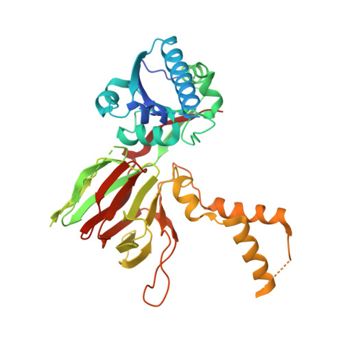
Crystal structure of human NADK2 reveals a dimeric organization and active site occlusion by lysine acetylation.

Mary, C.Soflaee, M.H.Kesavan, R.Gelin, M.Brown, H.Zacharias, G.Mathews, T.P.Lemoff, A.Lionne, C.Labesse, G.Hoxhaj, G.

(2022) Mol Cell 82: 3299

  • DOI: https://doi.org/10.1016/j.molcel.2022.06.026
  • Primary Citation of Related Structures:  
    7R4J, 7R4K, 7R4L, 7R4M

  • PubMed Abstract: 

    NAD + kinases (NADKs) are metabolite kinases that phosphorylate NAD + molecules to make NADP + , a limiting substrate for the generation of reducing power NADPH. NADK2 sustains mitochondrial NADPH production that enables proline biosynthesis and antioxidant defense. However, its molecular architecture and mechanistic regulation remain undescribed. Here, we report the crystal structure of human NADK2, revealing a substrate-driven mode of activation. We find that NADK2 presents an unexpected dimeric organization instead of the typical tetrameric assemblage observed for other NADKs. A specific extended segment (aa 325-365) is crucial for NADK2 dimerization and activity. Moreover, we characterize numerous acetylation events, including those on Lys76 and Lys304, which reside near the active site and inhibit NADK2 activity without disrupting dimerization, thereby reducing mitochondrial NADP(H) production, proline synthesis, and cell growth. These findings reveal important molecular insight into the structure and regulation of a vital enzyme in mitochondrial NADPH and proline metabolism.


  • Organizational Affiliation

    Atelier de Biologie Chimie Informatique Structurale, CNRS, INSERM, Univ Montpellier, Centre de Biologie Structurale, Montpellier, France.


Macromolecules
Find similar proteins by:  (by identity cutoff)  |  3D Structure
Entity ID: 1
MoleculeChains Sequence LengthOrganismDetailsImage
NAD kinase 2, mitochondrial391Homo sapiensMutation(s): 0 
Gene Names: NADK2C5orf33MNADKNADKD1
EC: 2.7.1.23
UniProt & NIH Common Fund Data Resources
Find proteins for Q4G0N4 (Homo sapiens)
Explore Q4G0N4 
Go to UniProtKB:  Q4G0N4
PHAROS:  Q4G0N4
GTEx:  ENSG00000152620 
Entity Groups  
Sequence Clusters30% Identity50% Identity70% Identity90% Identity95% Identity100% Identity
UniProt GroupQ4G0N4
Sequence Annotations
Expand
  • Reference Sequence
Small Molecules
Experimental Data & Validation

Experimental Data

  • Method: X-RAY DIFFRACTION
  • Resolution: 3.33 Å
  • R-Value Free: 0.287 
  • R-Value Work: 0.214 
  • R-Value Observed: 0.217 
  • Space Group: P 43 21 2
Unit Cell:
Length ( Å )Angle ( ˚ )
a = 67.583α = 90
b = 67.583β = 90
c = 221.815γ = 90
Software Package:
Software NamePurpose
PHENIXrefinement
XSCALEdata scaling
PHASERphasing
PDB_EXTRACTdata extraction
XDSdata reduction

Structure Validation

View Full Validation Report


Ligand Structure Quality Assessment 


Entry History & Funding Information

Deposition Data


Funding OrganizationLocationGrant Number
Agence Nationale de la Recherche (ANR)FranceANR-17-CE18-0011-01

Revision History  (Full details and data files)

  • Version 1.0: 2022-07-06
    Type: Initial release
  • Version 1.1: 2022-08-03
    Changes: Database references
  • Version 1.2: 2022-09-14
    Changes: Database references
  • Version 1.3: 2024-05-01
    Changes: Data collection, Refinement description