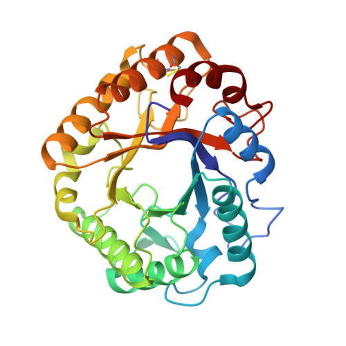
Definition of the catalytic cleft and product profile of a GH5_5 cellulase from Thermoascus aurantiacus.
Collet, L., Denys, A., Oudjama, Y., Sifennasr, K., Vander Wauven, C., Dutoit, R.(2026) Acta Crystallogr D Struct Biol
- PubMed: 42101894 Search on PubMed
- DOI: https://doi.org/10.1107/S2059798326003578
- Primary Citation Related Structures:
7R28, 7R29, 7R2A, 7R2C, 7R2D, 9F0N - PubMed Abstract:
Glycoside hydrolases (GHs) achieve glycan breakdown through glycosidic bond hydrolysis. Some retaining GHs can also transglycosylate, which could be useful for glycosynthesis. Improving GHs for either glycan degradation or synthesis requires a deep understanding of the residues involved in catalysis and substrate binding. This study characterizes in detail the activity and structure of Ta_Cel5A, a cellulase in glycoside hydrolase family 5, subfamily 5 (GH5_5) from the thermophilic ascomycete Thermoascus aurantiacus. While its hydrolytic activity was confirmed, Ta_Cel5A was also found to exhibit a weak transglycosylase activity with cellopentaose as a substrate. Transglycosylation products were detected within the first minutes of reaction at 25°C, far below its optimal temperature. The structures of catalytically impaired variants were solved in complex with oligosaccharides. The entire catalytic cleft was defined, consisting of seven glucose-binding subsites, five negative subsites and two positive subsites, from the nonreducing end to the reducing end. The fifth negative subsite could not be inferred in silico, showing the limitation in predicting distal subsites based on structural analogy. The structure of the glycosyl-enzyme intermediate was also obtained, revealing the displacement of key residues in the active site. The covalent binding of a glycosidic molecule triggers a major displacement of the nucleophilic residue, Glu244, changing its interaction network. The acid/base residue, Glu133, and a conserved tyrosine residue, Tyr201, are also displaced during glycosyl-enzyme intermediate formation, hinting at their role in the deglycosylation step.
- LABIRIS, 1 Avenue Emile Gryzon, 1070 Brussels, Belgium.
Organizational Affiliation: