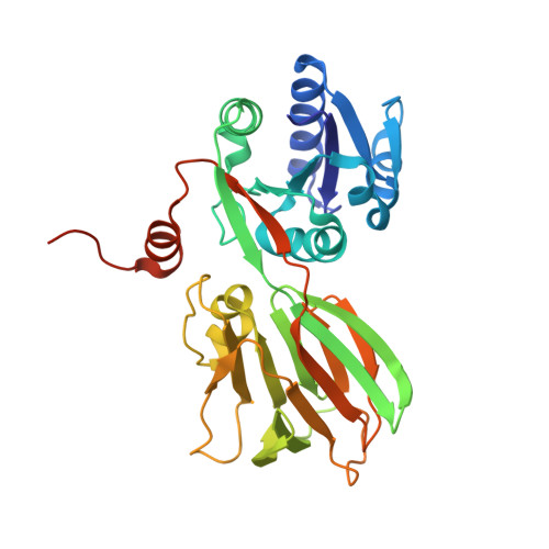
7QVS

Pseudomonas aeruginosa nicotinamide adenine dinucleotide kinase (NADK) structure in complex with NADP


Experimental Data Snapshot

  • Method: X-RAY DIFFRACTION
  • Resolution: 2.30 Å
  • R-Value Free: 0.236 
  • R-Value Work: 0.197 
  • R-Value Observed: 0.199 

wwPDB Validation   3D Report Full Report


Ligand Structure Quality Assessment 


This is version 1.2 of the entry. See complete history


Literature

Structure-based design, synthesis and biological evaluation of a NAD + analogue targeting Pseudomonas aeruginosa NAD kinase.

Rahimova, R.Nogaret, P.Huteau, V.Gelin, M.Clement, D.A.Labesse, G.Pochet, S.Blanc-Potard, A.B.Lionne, C.

(2023) FEBS J 290: 482-501

  • DOI: https://doi.org/10.1111/febs.16604
  • Primary Citation of Related Structures:  
    7QVS

  • PubMed Abstract: 

    Multidrug resistance is a major public health problem that requires the urgent development of new antibiotics and therefore the identification of novel bacterial targets. The activity of nicotinamide adenine dinucleotide kinase, NADK, is essential in all bacteria tested so far, including many human pathogens that display antibiotic resistance leading to the failure of current treatments. Inhibiting NADK is therefore a promising and innovative antibacterial strategy since there is currently no drug on the market targeting this enzyme. Through a fragment-based drug design approach, we have recently developed a NAD + -competitive inhibitor of NADKs, which displayed in vivo activity against Staphylococcus aureus. Here, we show that this compound, a di-adenosine derivative, is inactive against the NADK enzyme from the Gram-negative bacteria Pseudomonas aeruginosa (PaNADK). This lack of activity can be explained by the crystal structure of PaNADK, which was determined in complex with NADP + in this study. Structural analysis led us to design and synthesize a benzamide adenine dinucleoside analogue, active against PaNADK. This novel compound efficiently inhibited PaNADK enzymatic activity in vitro with a K i of 4.6 μm. Moreover, this compound reduced P. aeruginosa infection in vivo in a zebrafish model.


  • Organizational Affiliation

    Centre de Biologie Structurale (CBS), Université de Montpellier, CNRS UMR 5048, INSERM U1054, France.


Macromolecules
Find similar proteins by:  (by identity cutoff)  |  3D Structure
Entity ID: 1
MoleculeChains Sequence LengthOrganismDetailsImage
NAD kinase
A, B
303Pseudomonas aeruginosaMutation(s): 0 
Gene Names: 
EC: 2.7.1.23
UniProt
Find proteins for Q9HZC0 (Pseudomonas aeruginosa (strain ATCC 15692 / DSM 22644 / CIP 104116 / JCM 14847 / LMG 12228 / 1C / PRS 101 / PAO1))
Explore Q9HZC0 
Go to UniProtKB:  Q9HZC0
Entity Groups  
Sequence Clusters30% Identity50% Identity70% Identity90% Identity95% Identity100% Identity
UniProt GroupQ9HZC0
Sequence Annotations
Expand
  • Reference Sequence
Experimental Data & Validation

Experimental Data

  • Method: X-RAY DIFFRACTION
  • Resolution: 2.30 Å
  • R-Value Free: 0.236 
  • R-Value Work: 0.197 
  • R-Value Observed: 0.199 
  • Space Group: P 41 2 2
Unit Cell:
Length ( Å )Angle ( ˚ )
a = 88.77α = 90
b = 88.77β = 90
c = 174.247γ = 90
Software Package:
Software NamePurpose
PHENIXrefinement
PHENIXrefinement
Aimlessdata scaling
PDB_EXTRACTdata extraction
autoPROCdata reduction
PHASERphasing

Structure Validation

View Full Validation Report


Ligand Structure Quality Assessment 


Entry History & Funding Information

Deposition Data


Funding OrganizationLocationGrant Number
Agence Nationale de la Recherche (ANR)FranceANR-17-CE18-0011-02

Revision History  (Full details and data files)

  • Version 1.0: 2022-09-14
    Type: Initial release
  • Version 1.1: 2023-02-01
    Changes: Database references
  • Version 1.2: 2024-02-07
    Changes: Data collection, Refinement description