


Structural and Enzymological Characterization of Phosphoserine Phosphatase From Brucella melitensis
Pierson, E., Wouters, J.(2025) Proteins
Experimental Data Snapshot
Starting Model: in silico
View more details
wwPDB Validation 3D Report Full Report

(2025) Proteins
Entity ID: 1 | |||||
|---|---|---|---|---|---|
| Molecule | Chains | Sequence Length | Organism | Details | Image |
| O-phosphoserine phosphohydrolase | 307 | Brucella melitensis bv. 1 str. 16M | Mutation(s): 0 Gene Names: BMEI0615 EC: 3.1.3.3 | 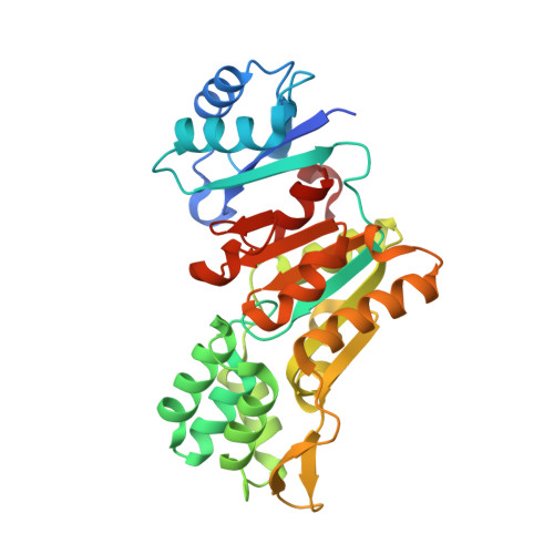 | |
UniProt | |||||
Entity Groups | |||||
| Sequence Clusters | 30% Identity50% Identity70% Identity90% Identity95% Identity100% Identity | ||||
| UniProt Group | Q8YI30 | ||||
Sequence AnnotationsExpand | |||||
Reference Sequence | |||||
| Ligands 6 Unique | |||||
|---|---|---|---|---|---|
| ID | Chains | Name / Formula / InChI Key | 2D Diagram | 3D Interactions | |
| PEG Download:Ideal Coordinates CCD File | B [auth A] | DI(HYDROXYETHYL)ETHER C4 H10 O3 MTHSVFCYNBDYFN-UHFFFAOYSA-N |  | ||
| PO4 Download:Ideal Coordinates CCD File | Q [auth A] | PHOSPHATE ION O4 P NBIIXXVUZAFLBC-UHFFFAOYSA-K |  | ||
| GOL Download:Ideal Coordinates CCD File | C [auth A] E [auth A] I [auth A] J [auth A] K [auth A] | GLYCEROL C3 H8 O3 PEDCQBHIVMGVHV-UHFFFAOYSA-N |  | ||
| EDO Download:Ideal Coordinates CCD File | L [auth A], M [auth A] | 1,2-ETHANEDIOL C2 H6 O2 LYCAIKOWRPUZTN-UHFFFAOYSA-N |  | ||
| FMT Download:Ideal Coordinates CCD File | D [auth A], F [auth A], G [auth A], H [auth A], O [auth A] | FORMIC ACID C H2 O2 BDAGIHXWWSANSR-UHFFFAOYSA-N |  | ||
| MG (Subject of Investigation/LOI) Download:Ideal Coordinates CCD File | P [auth A] | MAGNESIUM ION Mg JLVVSXFLKOJNIY-UHFFFAOYSA-N |  | ||
| Length ( Å ) | Angle ( ˚ ) |
|---|---|
| a = 143.21 | α = 90 |
| b = 143.21 | β = 90 |
| c = 143.21 | γ = 90 |
| Software Name | Purpose |
|---|---|
| BUSTER | refinement |
| PHENIX | refinement |
| MxCuBE | data collection |
| autoPROC | data processing |
| PHASER | phasing |
| Coot | model building |
| XDS | data reduction |
| XSCALE | data scaling |
| Funding Organization | Location | Grant Number |
|---|---|---|
| Other government | Belgium | -- |


