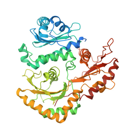
7QLA | pdb_00007qla

Structure of the Rab GEF complex Mon1-Ccz1


Experimental Data Snapshot

  • Method: ELECTRON MICROSCOPY
  • Resolution: 3.85 Å
  • Aggregation State: PARTICLE 
  • Reconstruction Method: SINGLE PARTICLE 

wwPDB Validation 3D Report Full Report

Validation slider image for 7QLA

This is version 1.3 of the entry. See complete history. 

Literature

Structure of the Mon1-Ccz1 complex reveals molecular basis of membrane binding for Rab7 activation.

Klink, B.U., Herrmann, E., Antoni, C., Langemeyer, L., Kiontke, S., Gatsogiannis, C., Ungermann, C., Raunser, S., Kummel, D.

(2022) Proc Natl Acad Sci U S A 119

  • DOI: https://doi.org/10.1073/pnas.2121494119
  • Primary Citation Related Structures: 
    7QLA

  • PubMed Abstract: 

    Activation of the GTPase Rab7/Ypt7 by its cognate guanine nucleotide exchange factor (GEF) Mon1-Ccz1 marks organelles such as endosomes and autophagosomes for fusion with lysosomes/vacuoles and degradation of their content. Here, we present a high-resolution cryogenic electron microscopy structure of the Mon1-Ccz1 complex that reveals its architecture in atomic detail. Mon1 and Ccz1 are arranged side by side in a pseudo-twofold symmetrical heterodimer. The three Longin domains of each Mon1 and Ccz1 are triangularly arranged, providing a strong scaffold for the catalytic center of the GEF. At the opposite side of the Ypt7-binding site, a positively charged and relatively flat patch stretches the Longin domains 2/3 of Mon1 and functions as a phosphatidylinositol phosphate-binding site, explaining how the GEF is targeted to membranes. Our work provides molecular insight into the mechanisms of endosomal Rab activation and serves as a blueprint for understanding the function of members of the Tri Longin domain Rab-GEF family.


  • Organizational Affiliation: 
    • Department of Structural Biochemistry, Max Planck Institute of Molecular Physiology, 44227 Dortmund, Germany.

Macromolecule Content 

  • Total Structure Weight: 133.35 kDa 
  • Atom Count: 6,837 
  • Modeled Residue Count: 874 
  • Deposited Residue Count: 1,221 
  • Unique protein chains: 2

Macromolecules

Find similar proteins by:|  3D Structure
Entity ID: 1
MoleculeChains  Sequence LengthOrganismDetailsImage
Vacuolar fusion protein MON1525Thermochaetoides thermophilaMutation(s): 0 
Gene Names: CTHT_0067370
UniProt
Find proteins for G0SGS3 (Thermochaetoides thermophila (strain DSM 1495 / CBS 144.50 / DAOM 24625 / IMI 039719))
Explore G0SGS3 
Go to UniProtKB:  G0SGS3
Entity Groups
Sequence Clusters30% Identity50% Identity70% Identity90% Identity95% Identity100% Identity
UniProt GroupG0SGS3
Sequence Annotations
Expand
Reference Sequence
Find similar proteins by:|  3D Structure
Entity ID: 2
MoleculeChains  Sequence LengthOrganismDetailsImage
Ccz1B [auth C]696Thermochaetoides thermophilaMutation(s): 0 
Gene Names: CTHT_0067370
UniProt
Find proteins for G0SD94 (Thermochaetoides thermophila (strain DSM 1495 / CBS 144.50 / DAOM 24625 / IMI 039719))
Explore G0SD94 
Go to UniProtKB:  G0SD94
Entity Groups
Sequence Clusters30% Identity50% Identity70% Identity90% Identity95% Identity100% Identity
UniProt GroupG0SD94
Sequence Annotations
Expand
Reference Sequence

Experimental Data & Validation

Experimental Data

  • Method: ELECTRON MICROSCOPY
  • Resolution: 3.85 Å
  • Aggregation State: PARTICLE 
  • Reconstruction Method: SINGLE PARTICLE 
EM Software:
TaskSoftware PackageVersion
MODEL REFINEMENTCoot
MODEL REFINEMENTPHENIX

Structure Validation

View Full Validation Report


Entry History 

& Funding Information

Deposition Data


Funding OrganizationLocationGrant Number
German Research Foundation (DFG)GermanySFB944-P17
Max Planck SocietyGermany--

Revision History  (Full details and data files)

  • Version 1.0: 2022-02-09
    Type: Initial release
  • Version 1.1: 2022-02-16
    Changes: Database references
  • Version 1.2: 2024-07-17
    Changes: Data collection
  • Version 1.3: 2025-07-09
    Changes: Data collection, Structure summary