H 2 S biogenesis by cystathionine beta-synthase: mechanism of inhibition by aminooxyacetic acid and unexpected role of serine.
Petrosino, M., Zuhra, K., Kopec, J., Hutchin, A., Szabo, C., Majtan, T.(2022) Cell Mol Life Sci 79: 438-438
- PubMed: 35864237 Search on PubMedSearch on PubMed Central
- DOI: https://doi.org/10.1007/s00018-022-04479-9
- Primary Citation Related Structures:
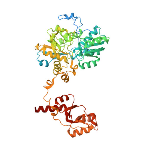
7QGT - PubMed Abstract:
Cystathionine beta-synthase (CBS) is a pivotal enzyme of the transsulfuration pathway responsible for diverting homocysteine to the biosynthesis of cysteine and production of hydrogen sulfide (H 2 S). Aberrant upregulation of CBS and overproduction of H 2 S contribute to pathophysiology of several diseases including cancer and Down syndrome. Therefore, pharmacological CBS inhibition has emerged as a prospective therapeutic approach. Here, we characterized binding and inhibitory mechanism of aminooxyacetic acid (AOAA), the most commonly used CBS inhibitor. We found that AOAA binds CBS tighter than its respective substrates and forms a dead-end PLP-bound intermediate featuring an oxime bond. Surprisingly, serine, but not cysteine, replaced AOAA from CBS and formed an aminoacrylate reaction intermediate, which allowed for the continuation of the catalytic cycle. Indeed, serine rescued and essentially normalized the enzymatic activity of AOAA-inhibited CBS. Cellular studies confirmed that AOAA decreased H 2 S production and bioenergetics, while additional serine rescued CBS activity, H 2 S production and mitochondrial function. The crystal structure of AOAA-bound human CBS showed a lack of hydrogen bonding with residues G305 and Y308, found in the serine-bound model. Thus, AOAA-inhibited CBS could be reactivated by serine. This difference may be important in a cellular environment in multiple pathophysiological conditions and may modulate the CBS-inhibitory activity of AOAA. In addition, our results demonstrate additional complexities of using AOAA as a CBS-specific inhibitor of H 2 S biogenesis and point to the urgent need to develop a potent, selective and specific pharmacological CBS inhibitor.
- Department of Pharmacology, Faculty of Science and Medicine, University of Fribourg, Chemin du Musee 18, PER17, 1700, Fribourg, Switzerland.
Organizational Affiliation: