7QB9

Structure of the ligand-free GPCR dimer Ste2


Experimental Data Snapshot

  • Method: ELECTRON MICROSCOPY
  • Resolution: 3.10 Å
  • Aggregation State: PARTICLE 
  • Reconstruction Method: SINGLE PARTICLE 

wwPDB Validation   3D Report Full Report


This is version 1.2 of the entry. See complete history


Literature

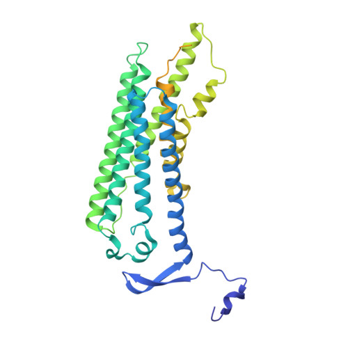
Activation mechanism of the class D fungal GPCR dimer Ste2.

Velazhahan, V.Ma, N.Vaidehi, N.Tate, C.G.

(2022) Nature 603: 743-748

  • DOI: https://doi.org/10.1038/s41586-022-04498-3
  • Primary Citation of Related Structures:  
    7QA8, 7QB9, 7QBC, 7QBI

  • PubMed Abstract: 

    The fungal class D1 G-protein-coupled receptor (GPCR) Ste2 has a different arrangement of transmembrane helices compared with mammalian GPCRs and a distinct mode of coupling to the heterotrimeric G protein Gpa1-Ste2-Ste18 1 . In addition, Ste2 lacks conserved sequence motifs such as DRY, PIF and NPXXY, which are associated with the activation of class A GPCRs 2 . This suggested that the activation mechanism of Ste2 may also differ. Here we determined structures of Saccharomyces cerevisiae Ste2 in the absence of G protein in two different conformations bound to the native agonist α-factor, bound to an antagonist and without ligand. These structures revealed that Ste2 is indeed activated differently from other GPCRs. In the inactive state, the cytoplasmic end of transmembrane helix H7 is unstructured and packs between helices H1-H6, blocking the G protein coupling site. Agonist binding results in the outward movement of the extracellular ends of H6 and H7 by 6 Å. On the intracellular surface, the G protein coupling site is formed by a 20 Å outward movement of the unstructured region in H7 that unblocks the site, and a 12 Å inward movement of H6. This is a distinct mechanism in GPCRs, in which the movement of H6 and H7 upon agonist binding facilitates G protein coupling.


  • Organizational Affiliation

    MRC Laboratory of Molecular Biology, Francis Crick Avenue, Cambridge, UK.


Macromolecules
Find similar proteins by:  (by identity cutoff)  |  3D Structure
Entity ID: 1
MoleculeChains Sequence LengthOrganismDetailsImage
Pheromone alpha factor receptor
A, B
431Saccharomyces cerevisiaeMutation(s): 0 
Gene Names: STE2
Membrane Entity: Yes 
UniProt
Find proteins for D6VTK4 (Saccharomyces cerevisiae (strain ATCC 204508 / S288c))
Explore D6VTK4 
Go to UniProtKB:  D6VTK4
Entity Groups  
Sequence Clusters30% Identity50% Identity70% Identity90% Identity95% Identity100% Identity
UniProt GroupD6VTK4
Sequence Annotations
Expand
  • Reference Sequence
Small Molecules
Ligands 2 Unique
IDChains Name / Formula / InChI Key2D Diagram3D Interactions
Y01
Query on Y01

Download Ideal Coordinates CCD File 
AA [auth B]
BA [auth B]
E [auth A]
F [auth A]
G [auth A]
AA [auth B],
BA [auth B],
E [auth A],
F [auth A],
G [auth A],
H [auth A],
I [auth A],
J [auth A],
K [auth A],
L [auth A],
M [auth A],
N [auth A],
O [auth A],
R [auth B],
S [auth B],
T [auth B],
U [auth B],
V [auth B],
W [auth B],
X [auth B],
Y [auth B],
Z [auth B]
CHOLESTEROL HEMISUCCINATE
C31 H50 O4
WLNARFZDISHUGS-MIXBDBMTSA-N
NAG
Query on NAG

Download Ideal Coordinates CCD File 
C [auth A],
D [auth A],
P [auth B],
Q [auth B]
2-acetamido-2-deoxy-beta-D-glucopyranose
C8 H15 N O6
OVRNDRQMDRJTHS-FMDGEEDCSA-N
Experimental Data & Validation

Experimental Data

  • Method: ELECTRON MICROSCOPY
  • Resolution: 3.10 Å
  • Aggregation State: PARTICLE 
  • Reconstruction Method: SINGLE PARTICLE 
EM Software:
TaskSoftware PackageVersion
RECONSTRUCTIONcryoSPARC3.1

Structure Validation

View Full Validation Report


Entry History & Funding Information

Deposition Data


Funding OrganizationLocationGrant Number
Medical Research Council (MRC, United Kingdom)United KingdomMRC U105197215

Revision History  (Full details and data files)

  • Version 1.0: 2022-03-16
    Type: Initial release
  • Version 1.1: 2022-03-30
    Changes: Database references
  • Version 1.2: 2022-04-06
    Changes: Database references