7Q9U

Crystal structure of the high affinity KRas mutant PDE6D complex


Experimental Data Snapshot

  • Method: X-RAY DIFFRACTION
  • Resolution: 2.24 Å
  • R-Value Free: 0.255 
  • R-Value Work: 0.199 

wwPDB Validation   3D Report Full Report


Ligand Structure Quality Assessment 


This is version 2.1 of the entry. See complete history


Literature

Stabilization of the RAS:PDE6D Complex Is a Novel Strategy to Inhibit RAS Signaling.

Yelland, T.Garcia, E.Parry, C.Kowalczyk, D.Wojnowska, M.Gohlke, A.Zalar, M.Cameron, K.Goodwin, G.Yu, Q.Zhu, P.C.ElMaghloob, Y.Pugliese, A.Archibald, L.Jamieson, A.Chen, Y.X.McArthur, D.Bower, J.Ismail, S.

(2022) J Med Chem 65: 1898-1914

  • DOI: https://doi.org/10.1021/acs.jmedchem.1c01265
  • Primary Citation of Related Structures:  
    7Q9Q, 7Q9R, 7Q9S, 7Q9U, 7QF9, 7QJK

  • PubMed Abstract: 

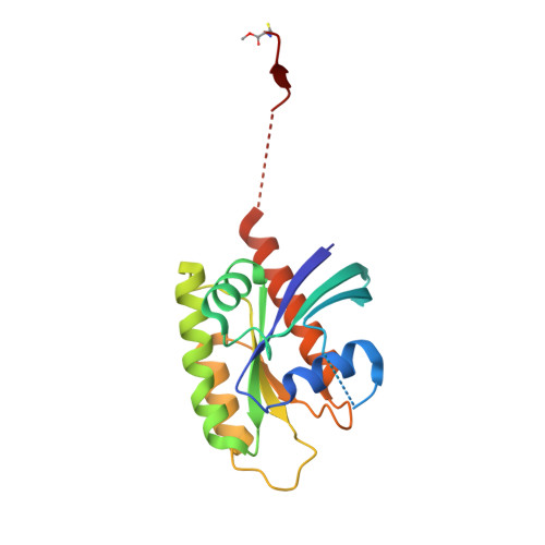
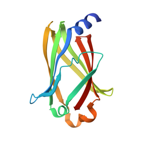
    RAS is a major anticancer drug target which requires membrane localization to activate downstream signal transduction. The direct inhibition of RAS has proven to be challenging. Here, we present a novel strategy for targeting RAS by stabilizing its interaction with the prenyl-binding protein PDE6D and disrupting its localization. Using rationally designed RAS point mutations, we were able to stabilize the RAS:PDE6D complex by increasing the affinity of RAS for PDE6D, which resulted in the redirection of RAS to the cytoplasm and the primary cilium and inhibition of oncogenic RAS/ERK signaling. We developed an SPR fragment screening and identified fragments that bind at the KRAS:PDE6D interface, as shown through cocrystal structures. Finally, we show that the stoichiometric ratios of KRAS:PDE6D vary in different cell lines, suggesting that the impact of this strategy might be cell-type-dependent. This study forms the foundation from which a potential anticancer small-molecule RAS:PDE6D complex stabilizer could be developed.


  • Organizational Affiliation

    CRUK Beatson Institute, Glasgow G61 1BD, United Kingdom.


Macromolecules
Find similar proteins by:  (by identity cutoff)  |  3D Structure
Entity ID: 1
MoleculeChains Sequence LengthOrganismDetailsImage
GTPase KRasA [auth AAA],
B [auth BBB]
185Homo sapiensMutation(s): 0 
Gene Names: KRASKRAS2RASK2
EC: 3.6.5.2
UniProt & NIH Common Fund Data Resources
Find proteins for P01116 (Homo sapiens)
Explore P01116 
Go to UniProtKB:  P01116
PHAROS:  P01116
GTEx:  ENSG00000133703 
Entity Groups  
Sequence Clusters30% Identity50% Identity70% Identity90% Identity95% Identity100% Identity
UniProt GroupP01116
Sequence Annotations
Expand
  • Reference Sequence
Find similar proteins by:  (by identity cutoff)  |  3D Structure
Entity ID: 2
MoleculeChains Sequence LengthOrganismDetailsImage
Retinal rod rhodopsin-sensitive cGMP 3',5'-cyclic phosphodiesterase subunit deltaC [auth DDD],
D [auth CCC]
150Homo sapiensMutation(s): 0 
Gene Names: PDE6DPDED
UniProt & NIH Common Fund Data Resources
Find proteins for O43924 (Homo sapiens)
Explore O43924 
Go to UniProtKB:  O43924
PHAROS:  O43924
GTEx:  ENSG00000156973 
Entity Groups  
Sequence Clusters30% Identity50% Identity70% Identity90% Identity95% Identity100% Identity
UniProt GroupO43924
Sequence Annotations
Expand
  • Reference Sequence
Small Molecules
Ligands 4 Unique
IDChains Name / Formula / InChI Key2D Diagram3D Interactions
GDP
Query on GDP

Download Ideal Coordinates CCD File 
E [auth AAA],
H [auth BBB]
GUANOSINE-5'-DIPHOSPHATE
C10 H15 N5 O11 P2
QGWNDRXFNXRZMB-UUOKFMHZSA-N
FAR
Query on FAR

Download Ideal Coordinates CCD File 
F [auth AAA],
I [auth BBB]
FARNESYL
C15 H26
JXBSHSBNOVLGHF-BUJBXKITSA-N
SO4
Query on SO4

Download Ideal Coordinates CCD File 
G [auth BBB],
J [auth DDD],
M [auth CCC]
SULFATE ION
O4 S
QAOWNCQODCNURD-UHFFFAOYSA-L
EDO
Query on EDO

Download Ideal Coordinates CCD File 
K [auth DDD],
L [auth DDD],
N [auth CCC],
O [auth CCC],
P [auth CCC]
1,2-ETHANEDIOL
C2 H6 O2
LYCAIKOWRPUZTN-UHFFFAOYSA-N
Modified Residues  1 Unique
IDChains TypeFormula2D DiagramParent
CMT
Query on CMT
A [auth AAA],
B [auth BBB]
L-PEPTIDE LINKINGC4 H9 N O2 SCYS
Experimental Data & Validation

Experimental Data

  • Method: X-RAY DIFFRACTION
  • Resolution: 2.24 Å
  • R-Value Free: 0.255 
  • R-Value Work: 0.199 
  • Space Group: P 1
Unit Cell:
Length ( Å )Angle ( ˚ )
a = 51.086α = 82.771
b = 57.925β = 81.709
c = 78.913γ = 68.447
Software Package:
Software NamePurpose
REFMACrefinement
DIALSdata reduction
Aimlessdata scaling
PHASERphasing

Structure Validation

View Full Validation Report


Ligand Structure Quality Assessment 


Entry History & Funding Information

Deposition Data


Funding OrganizationLocationGrant Number
Cancer Research UKUnited Kingdom--

Revision History  (Full details and data files)

  • Version 1.0: 2022-01-26
    Type: Initial release
  • Version 1.1: 2022-06-01
    Changes: Database references
  • Version 2.0: 2022-12-07
    Changes: Atomic model, Data collection, Database references, Derived calculations, Polymer sequence, Source and taxonomy, Structure summary
  • Version 2.1: 2024-01-31
    Changes: Data collection, Derived calculations, Refinement description