Crystal structures of WrbA, a spurious target of the salicylidene acylhydrazide inhibitors of type III secretion in Gram-negative pathogens, and verification of improved specificity of next-generation compounds.
Zambelloni, R., Beckham, K.S.H., Wu, H.J., Elofsson, M., Marquez, R., Gabrielsen, M., Roe, A.J.(2022) Microbiology (Reading) 168
- PubMed: 35829699 Search on PubMed
- DOI: https://doi.org/10.1099/mic.0.001211
- Primary Citation Related Structures:
7Q6M, 7Q6N, 7Q6O - PubMed Abstract:
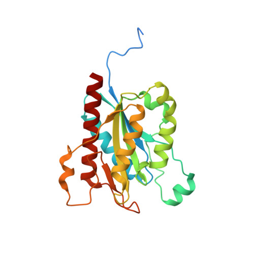
The enterohemorrhagic Escherichia coli pathotype is responsible for severe and dangerous infections in humans. Establishment of the infection requires colonization of the gastro-intestinal tract, which is dependent on the Type III Secretion System. The Type III Secretion System (T3SS) allows attachment of the pathogen to the mammalian host cell and cytoskeletal rearrangements within the host cell. Blocking the functionality of the T3SS is likely to reduce colonization and therefore limit the disease. This route offers an alternative to antibiotics, and problems with the development of antibiotics resistance. Salicylidene acylhydrazides have been shown to have an inhibitory effect on the T3SS in several pathogens. However, the main target of these compounds is still unclear. Past work has identified a number of putative protein targets of these compounds, one of which being WrbA. Whilst WrbA is considered an off-target interaction, this study presents the effect of the salicylidne acylhydrazide compounds on the activity of WrbA, along with crystal structures of WrbA from Yersinia pseudotuberculosis and Salmonella serovar Typhimurium; the latter also containing parts of the compound in the structure. We also present data showing that the original compounds were unstable in acidic conditions, and that later compounds showed improved stability.
- Institute of Infection, Immunity and Inflammation, College of Medical, Veterinary and Life Sciences, University of Glasgow, Glasgow, Glasgow, G12 8TA, UK.
Organizational Affiliation: