7Q4I | pdb_00007q4i

Crystal structure of DmC1GalT1 in complex with UDP-Mn2+ and the APD-TGalNAc-RP


Experimental Data Snapshot

  • Method: X-RAY DIFFRACTION
  • Resolution: 2.40 Å
  • R-Value Free: 
    0.235 (Depositor), 0.238 (DCC) 
  • R-Value Work: 
    0.175 (Depositor), 0.183 (DCC) 
  • R-Value Observed: 
    0.178 (Depositor) 

Starting Model: in silico
View more details

wwPDB Validation 3D Report Full Report

Validation slider image for 7Q4I

Ligand Structure Quality Assessment 


This is version 1.2 of the entry. See complete history

Literature

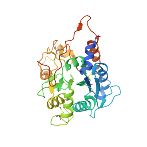
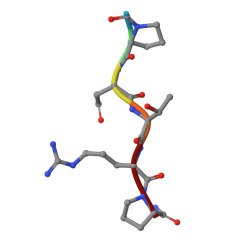
Structural basis for the synthesis of the core 1 structure by C1GalT1.

Gonzalez-Ramirez, A.M.Grosso, A.S.Yang, Z.Companon, I.Coelho, H.Narimatsu, Y.Clausen, H.Marcelo, F.Corzana, F.Hurtado-Guerrero, R.

(2022) Nat Commun 13: 2398-2398

  • DOI: https://doi.org/10.1038/s41467-022-29833-0
  • Primary Citation Related Structures: 
    7Q4I

  • PubMed Abstract: 

    C1GalT1 is an essential inverting glycosyltransferase responsible for synthesizing the core 1 structure, a common precursor for mucin-type O-glycans found in many glycoproteins. To date, the structure of C1GalT1 and the details of substrate recognition and catalysis remain unknown. Through biophysical and cellular studies, including X-ray crystallography of C1GalT1 complexed to a glycopeptide, we report that C1GalT1 is an obligate GT-A fold dimer that follows a S N 2 mechanism. The binding of the glycopeptides to the enzyme is mainly driven by the GalNAc moiety while the peptide sequence provides optimal kinetic and binding parameters. Interestingly, to achieve glycosylation, C1GalT1 recognizes a high-energy conformation of the α-GalNAc-Thr linkage, negligibly populated in solution. By imposing this 3D-arrangement on that fragment, characteristic of α-GalNAc-Ser peptides, C1GalT1 ensures broad glycosylation of both acceptor substrates. These findings illustrate a structural and mechanistic blueprint to explain glycosylation of multiple acceptor substrates, extending the repertoire of mechanisms adopted by glycosyltransferases.


  • Organizational Affiliation
    • Institute of Biocompuation and Physics of Complex Systems, University of Zaragoza, Mariano Esquillor s/n, Campus Rio Ebro, Edificio I+D, 50018, Zaragoza, Spain.

Macromolecule Content 

  • Total Structure Weight: 75.39 kDa 
  • Atom Count: 4,830 
  • Modeled Residue Count: 572 
  • Deposited Residue Count: 644 
  • Unique protein chains: 2

Macromolecules

Find similar proteins by:|  3D Structure
Entity ID: 1
MoleculeChains  Sequence LengthOrganismDetailsImage
Glycoprotein-N-acetylgalactosamine 3-beta-galactosyltransferase 1
A, B
316Drosophila melanogasterMutation(s): 0 
Gene Names: C1GalTACG9520
EC: 2.4.1.122
UniProt
Find proteins for Q7K237 (Drosophila melanogaster)
Explore Q7K237 
Go to UniProtKB:  Q7K237
Entity Groups
Sequence Clusters30% Identity50% Identity70% Identity90% Identity95% Identity100% Identity
UniProt GroupQ7K237
Sequence Annotations
Expand
Reference Sequence
Find similar proteins by:  Sequence   |   3D Structure  
Entity ID: 2
MoleculeChains  Sequence LengthOrganismDetailsImage
Mucin-1C [auth G],
D [auth F]
6Homo sapiensMutation(s): 0 
UniProt & NIH Common Fund Data Resources
Find proteins for P15941 (Homo sapiens)
Explore P15941 
Go to UniProtKB:  P15941
PHAROS:  P15941
GTEx:  ENSG00000185499 
Entity Groups
UniProt GroupP15941
Glycosylation
Glycosylation Sites: 1Go to GlyGen: P15941-1
Sequence Annotations
Expand
Reference Sequence

Small Molecules

Ligands 4 Unique
IDChains Name / Formula / InChI Key2D Diagram3D Interactions
UDP

Query on UDP


Download:Ideal Coordinates CCD File
E [auth A],
H [auth B]
URIDINE-5'-DIPHOSPHATE
C9 H14 N2 O12 P2
XCCTYIAWTASOJW-XVFCMESISA-N
A2G
(Subject of Investigation/LOI)

Query on A2G


Download:Ideal Coordinates CCD File
K [auth G],
M [auth F]
2-acetamido-2-deoxy-alpha-D-galactopyranose
C8 H15 N O6
OVRNDRQMDRJTHS-CBQIKETKSA-N
EDO

Query on EDO


Download:Ideal Coordinates CCD File
F [auth A],
I [auth B],
L [auth F]
1,2-ETHANEDIOL
C2 H6 O2
LYCAIKOWRPUZTN-UHFFFAOYSA-N
MN

Query on MN


Download:Ideal Coordinates CCD File
G [auth A],
J [auth B]
MANGANESE (II) ION
Mn
WAEMQWOKJMHJLA-UHFFFAOYSA-N
Modified Residues  1 Unique
IDChains TypeFormula2D DiagramParent
LPD
Query on LPD
C [auth G],
D [auth F]
L-PEPTIDE LINKINGC5 H10 N2 OPRO

Experimental Data & Validation

Experimental Data

  • Method: X-RAY DIFFRACTION
  • Resolution: 2.40 Å
  • R-Value Free:  0.235 (Depositor), 0.238 (DCC) 
  • R-Value Work:  0.175 (Depositor), 0.183 (DCC) 
  • R-Value Observed: 0.178 (Depositor) 
Space Group: P 1 21 1
Unit Cell:
Length ( Å )Angle ( ˚ )
a = 50.241α = 90
b = 80.445β = 93.19
c = 71.949γ = 90
Software Package:
Software NamePurpose
REFMACrefinement
XDSdata reduction
SCALAdata scaling
PHASERphasing

Structure Validation

View Full Validation Report


Ligand Structure Quality Assessment 


Entry History 

& Funding Information

Deposition Data


Funding OrganizationLocationGrant Number
Spanish Ministry of Science, Innovation, and UniversitiesSpainPID2019-105451GB-I00

Revision History  (Full details and data files)

  • Version 1.0: 2022-04-13
    Type: Initial release
  • Version 1.1: 2022-11-16
    Changes: Database references
  • Version 1.2: 2024-05-01
    Changes: Data collection, Refinement description