7Q1Y

X-ray structure of human A2ML1


Experimental Data Snapshot

  • Method: X-RAY DIFFRACTION
  • Resolution: 4.40 Å
  • R-Value Free: 0.276 
  • R-Value Work: 0.238 
  • R-Value Observed: 0.240 

wwPDB Validation   3D Report Full Report


This is version 1.2 of the entry. See complete history


Literature

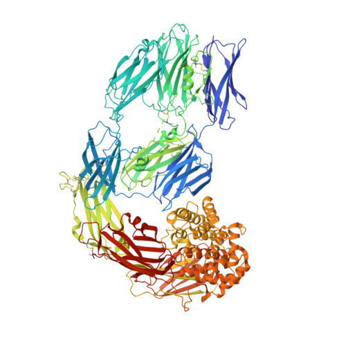
Cryo-EM structures of human A2ML1 elucidate the protease-inhibitory mechanism of the A2M family.

Nielsen, N.S.Zarantonello, A.Harwood, S.L.Jensen, K.T.Kjoge, K.Thogersen, I.B.Schauser, L.Karlsen, J.L.Andersen, G.R.Enghild, J.J.

(2022) Nat Commun 13: 3033-3033

  • DOI: https://doi.org/10.1038/s41467-022-30758-x
  • Primary Citation of Related Structures:  
    7Q1Y, 7Q5Z, 7Q60, 7Q61, 7Q62

  • PubMed Abstract: 

    A2ML1 is a monomeric protease inhibitor belonging to the A2M superfamily of protease inhibitors and complement factors. Here, we investigate the protease-inhibitory mechanism of human A2ML1 and determine the structures of its native and protease-cleaved conformations. The functional inhibitory unit of A2ML1 is a monomer that depends on covalent binding of the protease (mediated by A2ML1's thioester) to achieve inhibition. In contrast to the A2M tetramer which traps proteases in two internal chambers formed by four subunits, in protease-cleaved monomeric A2ML1 disordered regions surround the trapped protease and may prevent substrate access. In native A2ML1, the bait region is threaded through a hydrophobic channel, suggesting that disruption of this arrangement by bait region cleavage triggers the extensive conformational changes that result in protease inhibition. Structural comparisons with complement C3/C4 suggest that the A2M superfamily of proteins share this mechanism for the triggering of conformational change occurring upon proteolytic activation.


  • Organizational Affiliation

    Department of Molecular Biology and Genetics, Aarhus University, Aarhus, Denmark.


Macromolecules
Find similar proteins by:  (by identity cutoff)  |  3D Structure
Entity ID: 1
MoleculeChains Sequence LengthOrganismDetailsImage
Alpha-2-macroglobulin-like protein 1
A, B
1,436Homo sapiensMutation(s): 0 
Gene Names: A2ML1CPAMD9
UniProt & NIH Common Fund Data Resources
Find proteins for A8K2U0 (Homo sapiens)
Explore A8K2U0 
Go to UniProtKB:  A8K2U0
PHAROS:  A8K2U0
GTEx:  ENSG00000166535 
Entity Groups  
Sequence Clusters30% Identity50% Identity70% Identity90% Identity95% Identity100% Identity
UniProt GroupA8K2U0
Sequence Annotations
Expand
  • Reference Sequence
Oligosaccharides

Help

Entity ID: 2
MoleculeChains Length2D Diagram Glycosylation3D Interactions
beta-D-mannopyranose-(1-4)-2-acetamido-2-deoxy-beta-D-glucopyranose-(1-4)-2-acetamido-2-deoxy-beta-D-glucopyranose
C, D, E, F, I
3N-Glycosylation
Glycosylation Resources
GlyTouCan:  G15407YE
GlyCosmos:  G15407YE
GlyGen:  G15407YE
Entity ID: 3
MoleculeChains Length2D Diagram Glycosylation3D Interactions
2-acetamido-2-deoxy-beta-D-glucopyranose-(1-4)-2-acetamido-2-deoxy-beta-D-glucopyranose
G, H
2N-Glycosylation
Glycosylation Resources
GlyTouCan:  G42666HT
GlyCosmos:  G42666HT
GlyGen:  G42666HT
Experimental Data & Validation

Experimental Data

  • Method: X-RAY DIFFRACTION
  • Resolution: 4.40 Å
  • R-Value Free: 0.276 
  • R-Value Work: 0.238 
  • R-Value Observed: 0.240 
  • Space Group: H 3 2
Unit Cell:
Length ( Å )Angle ( ˚ )
a = 320.15α = 90
b = 320.15β = 90
c = 321.94γ = 120
Software Package:
Software NamePurpose
PHENIXrefinement
XDSdata reduction
XSCALEdata scaling
PHASERphasing

Structure Validation

View Full Validation Report


Entry History & Funding Information

Deposition Data


Funding OrganizationLocationGrant Number
LundbeckfondenDenmarkR155-2015-2666

Revision History  (Full details and data files)

  • Version 1.0: 2022-04-20
    Type: Initial release
  • Version 1.1: 2022-11-02
    Changes: Database references
  • Version 1.2: 2024-05-01
    Changes: Data collection, Refinement description