7PY4

Cryo-EM structure of ATP8B1-CDC50A in E2P autoinhibited state


Experimental Data Snapshot

  • Method: ELECTRON MICROSCOPY
  • Resolution: 3.10 Å
  • Aggregation State: PARTICLE 
  • Reconstruction Method: SINGLE PARTICLE 

wwPDB Validation   3D Report Full Report


This is version 1.0 of the entry. See complete history


Literature

Autoinhibition and regulation by phosphoinositides of ATP8B1, a human lipid flippase associated with intrahepatic cholestatic disorders.

Dieudonne, T.Herrera, S.A.Laursen, M.J.Lejeune, M.Stock, C.Slimani, K.Jaxel, C.Lyons, J.A.Montigny, C.Pomorski, T.G.Nissen, P.Lenoir, G.

(2022) Elife 11

  • DOI: https://doi.org/10.7554/eLife.75272
  • Primary Citation of Related Structures:  
    7PY4

  • PubMed Abstract: 

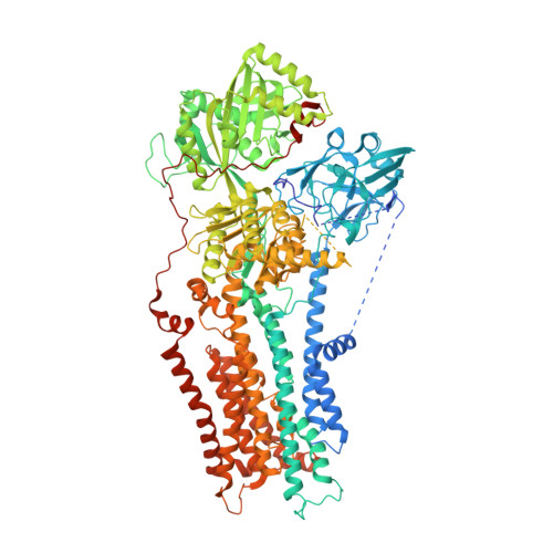
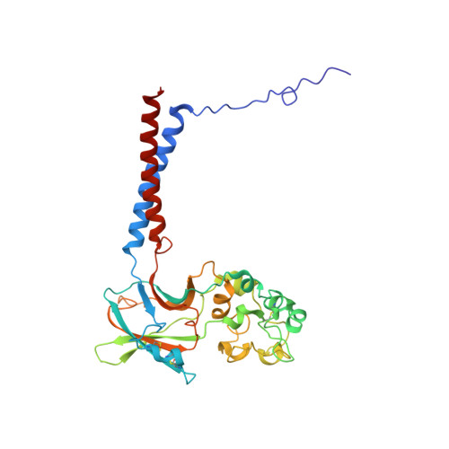
    P4-ATPases flip lipids from the exoplasmic to the cytosolic leaflet, thus maintaining lipid asymmetry in eukaryotic cell membranes. Mutations in several human P4-ATPase genes are associated with severe diseases, for example in ATP8B1 causing progressive familial intrahepatic cholestasis, a rare inherited disorder progressing toward liver failure. ATP8B1 forms a binary complex with CDC50A and displays a broad specificity to glycerophospholipids, but regulatory mechanisms are unknown. Here, we report functional studies and the cryo-EM structure of the human lipid flippase ATP8B1-CDC50A at 3.1 Å resolution. We find that ATP8B1 is autoinhibited by its N- and C-terminal tails, which form extensive interactions with the catalytic sites and flexible domain interfaces. Consistently, ATP hydrolysis is unleashed by truncation of the C-terminus, but also requires phosphoinositides, most markedly phosphatidylinositol-3,4,5-phosphate (PI(3,4,5)P 3 ), and removal of both N- and C-termini results in full activation. Restored inhibition of ATP8B1 truncation constructs with a synthetic peptide mimicking the C-terminal segment further suggests molecular communication between N- and C-termini in the autoinhibition and demonstrates that the regulatory mechanism can be interfered with by exogenous compounds. A recurring (G/A)(Y/F)AFS motif of the C-terminal segment suggests that this mechanism is employed widely across P4-ATPase lipid flippases in plasma membrane and endomembranes.


  • Organizational Affiliation

    Université Paris-Saclay, CEA, CNRS, Institute for Integrative Biology of the Cell (I2BC), Gif-sur-Yvette, France.


Macromolecules
Find similar proteins by:  (by identity cutoff)  |  3D Structure
Entity ID: 1
MoleculeChains Sequence LengthOrganismDetailsImage
Phospholipid-transporting ATPase IC1,256Homo sapiensMutation(s): 0 
Gene Names: ATP8B1ATPICFIC1PFIC
EC: 7.6.2.1
Membrane Entity: Yes 
UniProt & NIH Common Fund Data Resources
Find proteins for O43520 (Homo sapiens)
Explore O43520 
Go to UniProtKB:  O43520
PHAROS:  O43520
GTEx:  ENSG00000081923 
Entity Groups  
Sequence Clusters30% Identity50% Identity70% Identity90% Identity95% Identity100% Identity
UniProt GroupO43520
Sequence Annotations
Expand
  • Reference Sequence
Find similar proteins by:  (by identity cutoff)  |  3D Structure
Entity ID: 2
MoleculeChains Sequence LengthOrganismDetailsImage
Cell cycle control protein 50A327Homo sapiensMutation(s): 0 
Gene Names: TMEM30AC6orf67CDC50A
Membrane Entity: Yes 
UniProt & NIH Common Fund Data Resources
Find proteins for Q9NV96 (Homo sapiens)
Explore Q9NV96 
Go to UniProtKB:  Q9NV96
PHAROS:  Q9NV96
GTEx:  ENSG00000112697 
Entity Groups  
Sequence Clusters30% Identity50% Identity70% Identity90% Identity95% Identity100% Identity
UniProt GroupQ9NV96
Sequence Annotations
Expand
  • Reference Sequence
Oligosaccharides

Help

Entity ID: 3
MoleculeChains Length2D Diagram Glycosylation3D Interactions
beta-D-mannopyranose-(1-4)-2-acetamido-2-deoxy-beta-D-glucopyranose-(1-4)-2-acetamido-2-deoxy-beta-D-glucopyranose
C
3N-Glycosylation
Glycosylation Resources
GlyTouCan:  G15407YE
GlyCosmos:  G15407YE
GlyGen:  G15407YE
Small Molecules
Ligands 4 Unique
IDChains Name / Formula / InChI Key2D Diagram3D Interactions
Y01 (Subject of Investigation/LOI)
Query on Y01

Download Ideal Coordinates CCD File 
F [auth A],
G [auth A],
H [auth A],
I [auth B]
CHOLESTEROL HEMISUCCINATE
C31 H50 O4
WLNARFZDISHUGS-MIXBDBMTSA-N
NAG (Subject of Investigation/LOI)
Query on NAG

Download Ideal Coordinates CCD File 
J [auth B],
K [auth B]
2-acetamido-2-deoxy-beta-D-glucopyranose
C8 H15 N O6
OVRNDRQMDRJTHS-FMDGEEDCSA-N
BEF (Subject of Investigation/LOI)
Query on BEF

Download Ideal Coordinates CCD File 
E [auth A]BERYLLIUM TRIFLUORIDE ION
Be F3
OGIAHMCCNXDTIE-UHFFFAOYSA-K
MG (Subject of Investigation/LOI)
Query on MG

Download Ideal Coordinates CCD File 
D [auth A]MAGNESIUM ION
Mg
JLVVSXFLKOJNIY-UHFFFAOYSA-N
Experimental Data & Validation

Experimental Data

  • Method: ELECTRON MICROSCOPY
  • Resolution: 3.10 Å
  • Aggregation State: PARTICLE 
  • Reconstruction Method: SINGLE PARTICLE 
EM Software:
TaskSoftware PackageVersion
RECONSTRUCTIONcryoSPARC3
MODEL REFINEMENTPHENIX

Structure Validation

View Full Validation Report


Entry History & Funding Information

Deposition Data


Funding OrganizationLocationGrant Number
Agence Nationale de la Recherche (ANR)FranceANR-14-CE09-0022
H2020 Marie Curie Actions of the European CommissionFrance101024542
LundbeckfondenFranceR310-2018-3713
German Research Foundation (DFG)FranceGU 1133/11-1
European Molecular Biology Organization (EMBO)France7881
French Infrastructure for Integrated Structural Biology (FRISBI)FranceANR-10-INSB-05

Revision History  (Full details and data files)

  • Version 1.0: 2022-04-27
    Type: Initial release