Exiting the tunnel of uncertainty: crystal soak to validated hit.
Martin, M.P., Noble, M.E.M.(2022) Acta Crystallogr D Struct Biol 78: 1294-1302
- PubMed: 36322414 Search on PubMedSearch on PubMed Central
- DOI: https://doi.org/10.1107/S2059798322009986
- Primary Citation Related Structures:
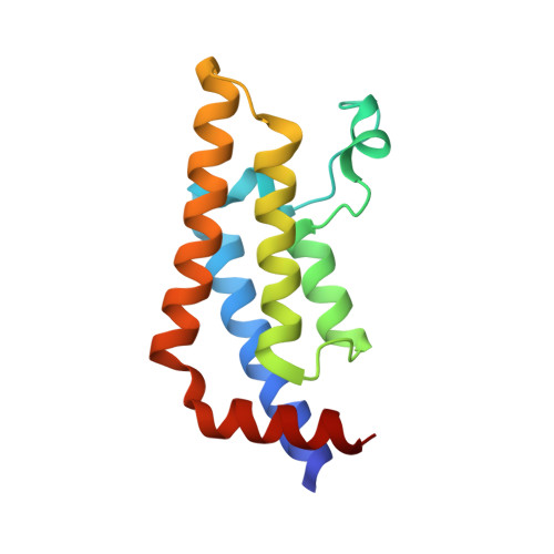
7PX5 - PubMed Abstract:
Crystallographic fragment screens provide an efficient and effective way to identify small-molecule ligands of a crystallized protein. Due to their low molecular weight, such hits tend to have low, often unquantifiable, affinity for their target, complicating the twin challenges of validating the hits as authentic solution-phase ligands of the target and identifying the `best' hit(s) for further elaboration. In this article, approaches that address these challenges are assessed. Using retrospective analysis of a recent ATAD2 hit-identification campaign, alongside other examples of successful fragment-screening campaigns, it is suggested that hit validation and prioritization are best achieved by a `triangulation' approach in which the results of multiple available biochemical and biophysical techniques are correlated to develop qualitative structure-activity relationships (SARs). Such qualitative SARs may indeed be the only means by which to navigate a project through the tunnel of uncertainty that prevails before on-scale biophysical, biochemical and/or biological measurements become possible.
- Cancer Research UK Drug Discovery Unit, Newcastle University, Paul O'Gorman Building, Framlington Place, Newcastle upon Tyne NE2 4HH, United Kingdom.
Organizational Affiliation: