7PS9 | pdb_00007ps9

Cereblon isoform 4 from Magnetospirillum gryphiswaldense in complex with Iberdomide (CC-220)


Experimental Data Snapshot

  • Method: X-RAY DIFFRACTION
  • Resolution: 1.80 Å
  • R-Value Free: 
    0.246 (Depositor), 0.244 (DCC) 
  • R-Value Work: 
    0.193 (Depositor), 0.200 (DCC) 
  • R-Value Observed: 
    0.196 (Depositor) 

Starting Model: experimental
View more details

wwPDB Validation 3D Report Full Report

Validation slider image for 7PS9

Ligand Structure Quality Assessment 


This is version 1.1 of the entry. See complete history

Literature

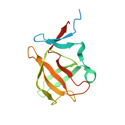
High-resolution structures of the bound effectors avadomide (CC-122) and iberdomide (CC-220) highlight advantages and limitations of the MsCI4 soaking system.

Heim, C.Hartmann, M.D.

(2022) Acta Crystallogr D Struct Biol 78: 290-298

  • DOI: https://doi.org/10.1107/S2059798322000092
  • Primary Citation Related Structures: 
    7PS9, 7PSO

  • PubMed Abstract: 

    Cereblon (CRBN) is the substrate receptor of the CRL4 CRBN E3 ubiquitin ligase and is a central player in targeted protein degradation. It is the target of the thalidomide-derived immunomodulatory drugs (IMiDs) and is one of the most widely employed receptors for proteolysis-targeting chimeras (PROTACs), both of which induce the ubiquitination and subsequent proteasomal degradation of target proteins. Structural studies of ligand binding to CRBN are crucial to elucidate the mechanisms of action and for mediation of side effects, ultimately aiding the development of next-generation IMiDs and PROTACs. With this aim, a crystal-soaking system based on the single-domain bacterial homologue MsCI4 has previously been established and used to delineate the binding modes of several classes of small molecules, including FDA-approved drugs, at the molecular level. Here, this system was used to characterize the binding of the next-generation IMiDs avadomide (CC-122) and iberdomide (CC-220) at high resolution, highlighting the advantages and limitations of the MsCI4 system and its implications for the development of future cereblon effectors.


  • Organizational Affiliation
    • Max Planck Institute for Developmental Biology, Max-Planck-Ring 5, 72076 Tübingen, Germany.

Macromolecule Content 

  • Total Structure Weight: 42.44 kDa 
  • Atom Count: 2,603 
  • Modeled Residue Count: 303 
  • Deposited Residue Count: 372 
  • Unique protein chains: 1

Macromolecules

Find similar proteins by:|  3D Structure
Entity ID: 1
MoleculeChains  Sequence LengthOrganismDetailsImage
Cereblon isoform 4
A, B, C
124Magnetospirillum gryphiswaldenseMutation(s): 0 
Gene Names: MGR_0879
UniProt
Find proteins for A4TVL0 (Magnetospirillum gryphiswaldense)
Explore A4TVL0 
Go to UniProtKB:  A4TVL0
Entity Groups
Sequence Clusters30% Identity50% Identity70% Identity90% Identity95% Identity100% Identity
UniProt GroupA4TVL0
Sequence Annotations
Expand
Reference Sequence

Experimental Data & Validation

Experimental Data

  • Method: X-RAY DIFFRACTION
  • Resolution: 1.80 Å
  • R-Value Free:  0.246 (Depositor), 0.244 (DCC) 
  • R-Value Work:  0.193 (Depositor), 0.200 (DCC) 
  • R-Value Observed: 0.196 (Depositor) 
Space Group: P 21 21 21
Unit Cell:
Length ( Å )Angle ( ˚ )
a = 56.001α = 90
b = 58.85β = 90
c = 88.583γ = 90
Software Package:
Software NamePurpose
REFMACrefinement
XDSdata reduction
XDSdata scaling
REFMACphasing

Structure Validation

View Full Validation Report


Ligand Structure Quality Assessment 


Entry History 

& Funding Information

Deposition Data


Funding OrganizationLocationGrant Number
Max Planck SocietyGermany--

Revision History  (Full details and data files)

  • Version 1.0: 2022-03-16
    Type: Initial release
  • Version 1.1: 2024-01-31
    Changes: Data collection, Refinement description