7PQP

tau-microtubule structural ensemble based on CryoEM data


Experimental Data Snapshot

  • Method: ELECTRON MICROSCOPY
  • Resolution: 4.10 Å
  • Aggregation State: FILAMENT 
  • Reconstruction Method: SINGLE PARTICLE 

wwPDB Validation   3D Report Full Report


This is version 1.1 of the entry. See complete history


Literature

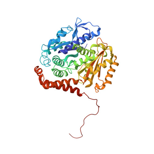
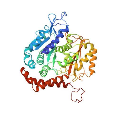
A Structural Ensemble of a Tau-Microtubule Complex Reveals Regulatory Tau Phosphorylation and Acetylation Mechanisms.

Brotzakis, Z.F.Lindstedt, P.R.Taylor, R.J.Rinauro, D.J.Gallagher, N.C.T.Bernardes, G.J.L.Vendruscolo, M.

(2021) ACS Cent Sci 7: 1986-1995

  • DOI: https://doi.org/10.1021/acscentsci.1c00585
  • Primary Citation of Related Structures:  
    7PQC, 7PQP

  • PubMed Abstract: 

    Tau is a microtubule-associated protein that regulates the stability of microtubules. We use metainference cryoelectron microscopy, an integrative structural biology approach, to determine an ensemble of conformations representing the structure and dynamics of a tau-microtubule complex comprising the entire microtubule-binding region of tau (residues 202-395). We thus identify the ground state of the complex and a series of excited states of lower populations. A comparison of the interactions in these different states reveals positions along the tau sequence that are important to determine the overall stability of the tau-microtubule complex. This analysis leads to the identification of positions where phosphorylation and acetylation events have destabilizing effects, which we validate by using site-specific post-translationally modified tau variants obtained by chemical mutagenesis. Taken together, these results illustrate how the simultaneous determination of ground and excited states of macromolecular complexes reveals functional and regulatory mechanisms.


  • Organizational Affiliation

    Centre for Misfolding Diseases, Department of Chemistry, University of Cambridge, Cambridge CB2 1EW, U.K.


Macromolecules
Find similar proteins by:  (by identity cutoff)  |  3D Structure
Entity ID: 1
MoleculeChains Sequence LengthOrganismDetailsImage
Tubulin beta chain
A, C, E, G, I
A, C, E, G, I, K, M
445Sus scrofaMutation(s): 0 
UniProt
Find proteins for P02554 (Sus scrofa)
Explore P02554 
Go to UniProtKB:  P02554
Entity Groups  
Sequence Clusters30% Identity50% Identity70% Identity90% Identity95% Identity100% Identity
UniProt GroupP02554
Sequence Annotations
Expand
  • Reference Sequence
Find similar proteins by:  (by identity cutoff)  |  3D Structure
Entity ID: 2
MoleculeChains Sequence LengthOrganismDetailsImage
Tubulin alpha-1B chain
B, D, F, H, J
B, D, F, H, J, L, N
451Sus scrofaMutation(s): 0 
Gene Names: TUBA1B
UniProt
Find proteins for Q2XVP4 (Sus scrofa)
Explore Q2XVP4 
Go to UniProtKB:  Q2XVP4
Entity Groups  
Sequence Clusters30% Identity50% Identity70% Identity90% Identity95% Identity100% Identity
UniProt GroupQ2XVP4
Sequence Annotations
Expand
  • Reference Sequence
Find similar proteins by:  (by identity cutoff)  |  3D Structure
Entity ID: 3
MoleculeChains Sequence LengthOrganismDetailsImage
Isoform Tau-F of Microtubule-associated protein tau194Homo sapiensMutation(s): 0 
Gene Names: MAPTMAPTLMTBT1TAU
UniProt & NIH Common Fund Data Resources
Find proteins for P10636 (Homo sapiens)
Explore P10636 
Go to UniProtKB:  P10636
PHAROS:  P10636
GTEx:  ENSG00000186868 
Entity Groups  
Sequence Clusters30% Identity50% Identity70% Identity90% Identity95% Identity100% Identity
UniProt GroupP10636
Sequence Annotations
Expand
  • Reference Sequence
Small Molecules
Ligands 3 Unique
IDChains Name / Formula / InChI Key2D Diagram3D Interactions
GTP (Subject of Investigation/LOI)
Query on GTP

Download Ideal Coordinates CCD File 
CA [auth J]
FA [auth L]
IA [auth N]
Q [auth B]
T [auth D]
CA [auth J],
FA [auth L],
IA [auth N],
Q [auth B],
T [auth D],
W [auth F],
Z [auth H]
GUANOSINE-5'-TRIPHOSPHATE
C10 H16 N5 O14 P3
XKMLYUALXHKNFT-UUOKFMHZSA-N
GDP (Subject of Investigation/LOI)
Query on GDP

Download Ideal Coordinates CCD File 
BA [auth I]
EA [auth K]
HA [auth M]
P [auth A]
S [auth C]
BA [auth I],
EA [auth K],
HA [auth M],
P [auth A],
S [auth C],
V [auth E],
Y [auth G]
GUANOSINE-5'-DIPHOSPHATE
C10 H15 N5 O11 P2
QGWNDRXFNXRZMB-UUOKFMHZSA-N
MG (Subject of Investigation/LOI)
Query on MG

Download Ideal Coordinates CCD File 
AA [auth H]
DA [auth J]
GA [auth L]
JA [auth N]
R [auth B]
AA [auth H],
DA [auth J],
GA [auth L],
JA [auth N],
R [auth B],
U [auth D],
X [auth F]
MAGNESIUM ION
Mg
JLVVSXFLKOJNIY-UHFFFAOYSA-N
Experimental Data & Validation

Experimental Data

  • Method: ELECTRON MICROSCOPY
  • Resolution: 4.10 Å
  • Aggregation State: FILAMENT 
  • Reconstruction Method: SINGLE PARTICLE 

Structure Validation

View Full Validation Report


Entry History & Funding Information

Deposition Data


Funding OrganizationLocationGrant Number
Royal SocietyUnited Kingdom--

Revision History  (Full details and data files)

  • Version 1.0: 2021-12-15
    Type: Initial release
  • Version 1.1: 2022-01-12
    Changes: Database references