7PQ9

Crystal structure of Bacillus clausii pdxR at 2.8 Angstroms resolution


Experimental Data Snapshot

  • Method: X-RAY DIFFRACTION
  • Resolution: 2.80 Å
  • R-Value Free: 0.254 
  • R-Value Work: 0.209 

wwPDB Validation   3D Report Full Report


This is version 1.2 of the entry. See complete history


Literature

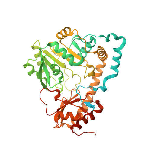
Structural insights into the DNA recognition mechanism by the bacterial transcription factor PdxR.

Freda, I.Exertier, C.Barile, A.Chaves-Sanjuan, A.Vega, M.V.Isupov, M.N.Harmer, N.J.Gugole, E.Swuec, P.Bolognesi, M.Scipioni, A.Savino, C.Di Salvo, M.L.Contestabile, R.Vallone, B.Tramonti, A.Montemiglio, L.C.

(2023) Nucleic Acids Res 51: 8237-8254

  • DOI: https://doi.org/10.1093/nar/gkad552
  • Primary Citation of Related Structures:  
    7PQ9, 7ZLA, 7ZN5, 7ZPA, 7ZTH

  • PubMed Abstract: 

    Specificity in protein-DNA recognition arises from the synergy of several factors that stem from the structural and chemical signatures encoded within the targeted DNA molecule. Here, we deciphered the nature of the interactions driving DNA recognition and binding by the bacterial transcription factor PdxR, a member of the MocR family responsible for the regulation of pyridoxal 5'-phosphate (PLP) biosynthesis. Single particle cryo-EM performed on the PLP-PdxR bound to its target DNA enabled the isolation of three conformers of the complex, which may be considered as snapshots of the binding process. Moreover, the resolution of an apo-PdxR crystallographic structure provided a detailed description of the transition of the effector domain to the holo-PdxR form triggered by the binding of the PLP effector molecule. Binding analyses of mutated DNA sequences using both wild type and PdxR variants revealed a central role of electrostatic interactions and of the intrinsic asymmetric bending of the DNA in allosterically guiding the holo-PdxR-DNA recognition process, from the first encounter through the fully bound state. Our results detail the structure and dynamics of the PdxR-DNA complex, clarifying the mechanism governing the DNA-binding mode of the holo-PdxR and the regulation features of the MocR family of transcription factors.


  • Organizational Affiliation

    Department of Biochemical Sciences "A. Rossi Fanelli", Sapienza, University of Rome, Rome 00185, Italy.


Macromolecules
Find similar proteins by:  (by identity cutoff)  |  3D Structure
Entity ID: 1
MoleculeChains Sequence LengthOrganismDetailsImage
PLP-dependent aminotransferase family protein478Shouchella clausiiMutation(s): 0 
Gene Names: HJ578_10545
Entity Groups  
Sequence Clusters30% Identity50% Identity70% Identity90% Identity95% Identity100% Identity
Sequence Annotations
Expand
  • Reference Sequence
Small Molecules
Ligands 6 Unique
IDChains Name / Formula / InChI Key2D Diagram3D Interactions
P6G
Query on P6G

Download Ideal Coordinates CCD File 
JA [auth HHH]HEXAETHYLENE GLYCOL
C12 H26 O7
IIRDTKBZINWQAW-UHFFFAOYSA-N
PGE
Query on PGE

Download Ideal Coordinates CCD File 
X [auth EEE]TRIETHYLENE GLYCOL
C6 H14 O4
ZIBGPFATKBEMQZ-UHFFFAOYSA-N
PEG
Query on PEG

Download Ideal Coordinates CCD File 
CA [auth GGG]DI(HYDROXYETHYL)ETHER
C4 H10 O3
MTHSVFCYNBDYFN-UHFFFAOYSA-N
EDO
Query on EDO

Download Ideal Coordinates CCD File 
BA [auth GGG]1,2-ETHANEDIOL
C2 H6 O2
LYCAIKOWRPUZTN-UHFFFAOYSA-N
CA
Query on CA

Download Ideal Coordinates CCD File 
AA [auth FFF]
DA [auth GGG]
EA [auth GGG]
FA [auth GGG]
GA [auth HHH]
AA [auth FFF],
DA [auth GGG],
EA [auth GGG],
FA [auth GGG],
GA [auth HHH],
HA [auth HHH],
IA [auth HHH],
KA [auth III],
L [auth AAA],
LA [auth III],
M [auth AAA],
MA [auth III],
NA [auth JJJ],
OA [auth JJJ],
P [auth BBB],
Q [auth BBB],
QA [auth KKK],
R [auth CCC],
S [auth CCC],
T [auth CCC],
U [auth DDD],
V [auth DDD],
W [auth DDD],
Y [auth EEE],
Z [auth EEE]
CALCIUM ION
Ca
BHPQYMZQTOCNFJ-UHFFFAOYSA-N
CL
Query on CL

Download Ideal Coordinates CCD File 
N [auth AAA],
O [auth AAA],
PA [auth JJJ]
CHLORIDE ION
Cl
VEXZGXHMUGYJMC-UHFFFAOYSA-M
Experimental Data & Validation

Experimental Data

  • Method: X-RAY DIFFRACTION
  • Resolution: 2.80 Å
  • R-Value Free: 0.254 
  • R-Value Work: 0.209 
  • Space Group: P 61 2 2
Unit Cell:
Length ( Å )Angle ( ˚ )
a = 250.84α = 90
b = 250.84β = 90
c = 370.3γ = 120
Software Package:
Software NamePurpose
REFMACrefinement
XDSdata reduction
XSCALEdata scaling
MoRDaphasing
PARROTphasing
DMphasing
Cootmodel building

Structure Validation

View Full Validation Report


Entry History & Funding Information

Deposition Data


Funding OrganizationLocationGrant Number
Defence Science and Technology Laboratory (DSTL)United KingdomDSTL/AGR/00210/01-R1000094053

Revision History  (Full details and data files)

  • Version 1.0: 2022-09-28
    Type: Initial release
  • Version 1.1: 2023-10-11
    Changes: Data collection, Database references, Refinement description
  • Version 1.2: 2024-01-31
    Changes: Refinement description