7PHT | pdb_00007pht

Structure of Insulin receptor's transmembrane domain


Experimental Data Snapshot

  • Method: SOLUTION NMR
  • Conformers Calculated: 100 
  • Conformers Submitted: 20 
  • Selection Criteria: target function 

wwPDB Validation 3D Report Full Report

Validation slider image for 7PHT

This is version 1.1 of the entry. See complete history

Literature

Structure of Insulin receptor's transmembrane domain

Bershatsky, Y.V.Nadezhdin, K.D.Bocharova, O.V.Bocharov, E.V.

To be published.

Macromolecule Content 

  • Total Structure Weight: 3.46 kDa 
  • Atom Count: 245 
  • Modeled Residue Count: 30 
  • Deposited Residue Count: 30 
  • Unique protein chains: 1

Macromolecules

Find similar proteins by:|  3D Structure
Entity ID: 1
MoleculeChains  Sequence LengthOrganismDetailsImage
Isoform Long of Insulin receptor30Homo sapiensMutation(s): 0 
Gene Names: INSR
EC: 2.7.10.1
Membrane Entity: Yes 
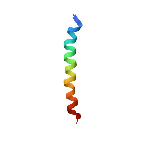
UniProt & NIH Common Fund Data Resources
Find proteins for P06213 (Homo sapiens)
Explore P06213 
Go to UniProtKB:  P06213
PHAROS:  P06213
GTEx:  ENSG00000171105 
Entity Groups
Sequence Clusters30% Identity50% Identity70% Identity90% Identity95% Identity100% Identity
UniProt GroupP06213
Sequence Annotations
Expand
Reference Sequence

Experimental Data & Validation

Experimental Data

  • Method: SOLUTION NMR
  • Conformers Calculated: 100 
  • Conformers Submitted: 20 
  • Selection Criteria: target function 

Structure Validation

View Full Validation Report


Entry History 

& Funding Information

Deposition Data


Funding OrganizationLocationGrant Number
Russian Science FoundationRussian Federation18-14-00375

Revision History  (Full details and data files)

  • Version 1.0: 2022-09-07
    Type: Initial release
  • Version 1.1: 2024-06-19
    Changes: Data collection, Database references