7PC7

The PDZ domain of SNTG1 complexed with the acetylated PDZ-binding motif of PTEN


Experimental Data Snapshot

  • Method: X-RAY DIFFRACTION
  • Resolution: 2.10 Å
  • R-Value Free: 0.218 
  • R-Value Work: 0.185 
  • R-Value Observed: 0.187 

wwPDB Validation   3D Report Full Report


This is version 1.1 of the entry. See complete history


Literature

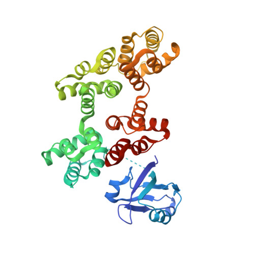
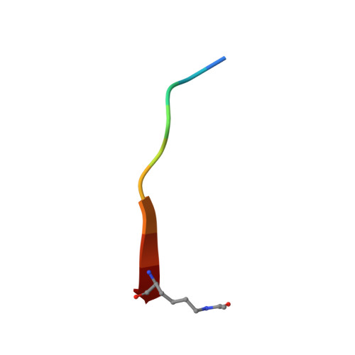
A scalable strategy to solve structures of PDZ domains and their complexes.

Cousido-Siah, A.Carneiro, L.Kostmann, C.Ecsedi, P.Nyitray, L.Trave, G.Gogl, G.

(2022) Acta Crystallogr D Struct Biol 78: 509-516

  • DOI: https://doi.org/10.1107/S2059798322001784
  • Primary Citation of Related Structures:  
    7PC3, 7PC4, 7PC5, 7PC7, 7PC8, 7PC9, 7PCB, 7QQL, 7QQM, 7QQN

  • PubMed Abstract: 

    The human PDZome represents one of the largest globular domain families in the human proteome, with 266 instances. These globular domains typically interact with C-terminal peptide motifs found in thousands of human proteins. Despite previous efforts, not all PDZ domains have experimentally solved structures and most of their complexes remain to be solved. Here, a simple and cost-effective strategy is proposed for the crystallization of PDZ domains and their complexes. A human annexin A2 fusion tag was used as a crystallization chaperone and the structures of nine PDZ domains were solved, including five domains that had not yet been solved. Finally, these novel experimental structures were compared with AlphaFold predictions and it is speculated how predictions and experimental methods could cooperate in order to investigate the structural landscapes of entire domain families and interactomes.


  • Organizational Affiliation

    Équipe Labellisée Ligue 2015, Département de Biologie Structurale Intégrative, Institut de Génétique et de Biologie Moléculaire et Cellulaire (IGBMC), INSERM U1258/CNRS UMR 7104/Université de Strasbourg, 1 Rue Laurent Fries, BP 10142, 67404 Illkirch, France.


Macromolecules
Find similar proteins by:  (by identity cutoff)  |  3D Structure
Entity ID: 1
MoleculeChains Sequence LengthOrganismDetailsImage
Gamma-1-syntrophin,Annexin A2
A, B
414Homo sapiensMutation(s): 0 
Gene Names: SNTG1ANXA2ANX2ANX2L4CAL1HLPC2D
UniProt & NIH Common Fund Data Resources
Find proteins for Q9NSN8 (Homo sapiens)
Explore Q9NSN8 
Go to UniProtKB:  Q9NSN8
PHAROS:  Q9NSN8
GTEx:  ENSG00000147481 
Find proteins for P07355 (Homo sapiens)
Explore P07355 
Go to UniProtKB:  P07355
PHAROS:  P07355
GTEx:  ENSG00000182718 
Entity Groups  
Sequence Clusters30% Identity50% Identity70% Identity90% Identity95% Identity100% Identity
UniProt GroupsP07355Q9NSN8
Sequence Annotations
Expand
  • Reference Sequence

Find similar proteins by:  Sequence   |   3D Structure  

Entity ID: 2
MoleculeChains Sequence LengthOrganismDetailsImage
Phosphatidylinositol 3,4,5-trisphosphate 3-phosphatase and dual-specificity protein phosphatase PTENC [auth E],
D [auth F]
10Homo sapiensMutation(s): 0 
EC: 3.1.3.16 (PDB Primary Data), 3.1.3.48 (PDB Primary Data), 3.1.3.67 (PDB Primary Data)
UniProt & NIH Common Fund Data Resources
Find proteins for P60484 (Homo sapiens)
Explore P60484 
Go to UniProtKB:  P60484
PHAROS:  P60484
GTEx:  ENSG00000171862 
Entity Groups  
Sequence Clusters30% Identity50% Identity70% Identity90% Identity95% Identity100% Identity
UniProt GroupP60484
Sequence Annotations
Expand
  • Reference Sequence
Small Molecules
Ligands 2 Unique
IDChains Name / Formula / InChI Key2D Diagram3D Interactions
GOL
Query on GOL

Download Ideal Coordinates CCD File 
J [auth A],
K [auth A],
R [auth B],
S [auth B]
GLYCEROL
C3 H8 O3
PEDCQBHIVMGVHV-UHFFFAOYSA-N
CA
Query on CA

Download Ideal Coordinates CCD File 
E [auth A]
F [auth A]
G [auth A]
H [auth A]
I [auth A]
E [auth A],
F [auth A],
G [auth A],
H [auth A],
I [auth A],
L [auth A],
M [auth B],
N [auth B],
O [auth B],
P [auth B],
Q [auth B],
T [auth B]
CALCIUM ION
Ca
BHPQYMZQTOCNFJ-UHFFFAOYSA-N
Modified Residues  1 Unique
IDChains TypeFormula2D DiagramParent
ALY
Query on ALY
C [auth E],
D [auth F]
L-PEPTIDE LINKINGC8 H16 N2 O3LYS
Experimental Data & Validation

Experimental Data

  • Method: X-RAY DIFFRACTION
  • Resolution: 2.10 Å
  • R-Value Free: 0.218 
  • R-Value Work: 0.185 
  • R-Value Observed: 0.187 
  • Space Group: P 21 21 21
Unit Cell:
Length ( Å )Angle ( ˚ )
a = 60.31α = 90
b = 61.74β = 90
c = 285.39γ = 90
Software Package:
Software NamePurpose
PHENIXrefinement
PDB_EXTRACTdata extraction
XDSdata reduction
XSCALEdata scaling
PHASERphasing

Structure Validation

View Full Validation Report


Entry History & Funding Information

Deposition Data


Funding OrganizationLocationGrant Number
Not funded--

Revision History  (Full details and data files)

  • Version 1.0: 2022-04-20
    Type: Initial release
  • Version 1.1: 2024-01-31
    Changes: Data collection, Refinement description