7PC2 | pdb_00007pc2

HIV-1 Env (BG505 SOSIP.664) in complex with the IgA bNAb 7-269 and the antibody 3BNC117.


Experimental Data Snapshot

  • Method: ELECTRON MICROSCOPY
  • Resolution: 2.80 Å
  • Aggregation State: PARTICLE 
  • Reconstruction Method: SINGLE PARTICLE 

Starting Model: experimental
View more details

wwPDB Validation 3D Report Full Report

Validation slider image for 7PC2

This is version 1.2 of the entry. See complete history

Literature

Epitope convergence of broadly HIV-1 neutralizing IgA and IgG antibody lineages in a viremic controller.

Lorin, V.Fernandez, I.Masse-Ranson, G.Bouvin-Pley, M.Molinos-Albert, L.M.Planchais, C.Hieu, T.Pehau-Arnaudet, G.Hrebik, D.Girelli-Zubani, G.Fiquet, O.Guivel-Benhassine, F.Sanders, R.W.Walker, B.D.Schwartz, O.Scheid, J.F.Dimitrov, J.D.Plevka, P.Braibant, M.Seaman, M.S.Bontems, F.Di Santo, J.P.Rey, F.A.Mouquet, H.

(2022) J Exp Medicine 219

  • DOI: https://doi.org/10.1084/jem.20212045
  • Primary Citation Related Structures: 
    7PC2

  • PubMed Abstract: 

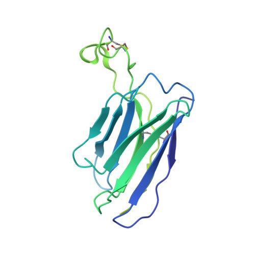
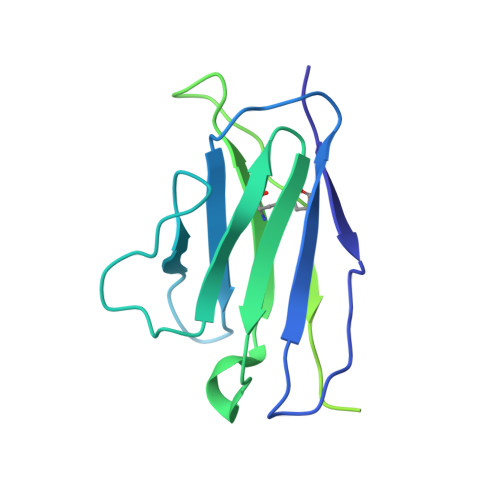
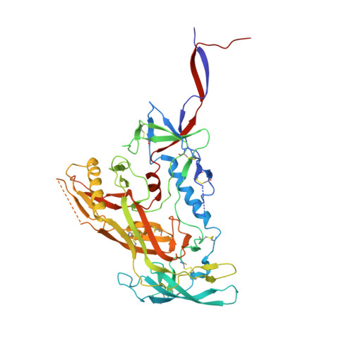
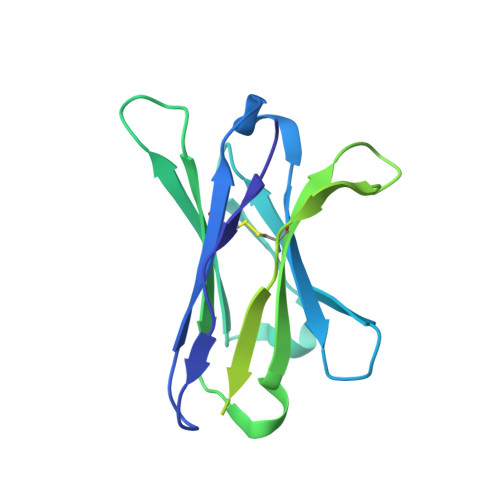
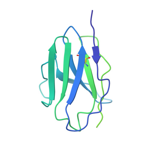
    Decrypting the B cell ontogeny of HIV-1 broadly neutralizing antibodies (bNAbs) is paramount for vaccine design. Here, we characterized IgA and IgG bNAbs of three distinct B cell lineages in a viremic controller, two of which comprised only IgG+ or IgA+ blood memory B cells; the third combined both IgG and IgA clonal variants. 7-269 bNAb in the IgA-only lineage displayed the highest neutralizing capacity despite limited somatic mutation, and delayed viral rebound in humanized mice. bNAbs in all three lineages targeted the N332 glycan supersite. The 2.8-Å resolution cryo-EM structure of 7-269-BG505 SOSIP.664 complex showed a similar pose as 2G12, on an epitope mainly composed of sugar residues comprising the N332 and N295 glycans. Binding and cryo-EM structural analyses showed that antibodies from the two other lineages interact mostly with glycans N332 and N386. Hence, multiple B cell lineages of IgG and IgA bNAbs focused on a unique HIV-1 site of vulnerability can codevelop in HIV-1 viremic controllers.


  • Organizational Affiliation
    • Laboratory of Humoral Immunology, Department of Immunology, Institut Pasteur, Paris, France.

Macromolecule Content 

  • Total Structure Weight: 546.14 kDa 
  • Atom Count: 27,063 
  • Modeled Residue Count: 3,093 
  • Deposited Residue Count: 4,563 
  • Unique protein chains: 6

Macromolecules

Find similar proteins by:|  3D Structure
Entity ID: 1
MoleculeChains  Sequence LengthOrganismDetailsImage
7-269 IgA Fab heavy chainA [auth O],
C [auth M],
E [auth Q]
240Homo sapiensMutation(s): 0 
Find similar proteins by:|  3D Structure
Entity ID: 2
MoleculeChains  Sequence LengthOrganismDetailsImage
7-269 IgA Fab light chainB [auth P],
D [auth N],
F [auth R]
215Homo sapiensMutation(s): 0 
Find similar proteins by:|  3D Structure
Entity ID: 3
MoleculeChains  Sequence LengthOrganismDetailsImage
gp120, BG505 SOSIP.664 T332NG [auth A],
H [auth B],
I [auth C]
481Human immunodeficiency virusMutation(s): 0 
Gene Names: env
UniProt
Find proteins for Q2N0S6 (Human immunodeficiency virus type 1)
Explore Q2N0S6 
Go to UniProtKB:  Q2N0S6
Entity Groups
Sequence Clusters30% Identity50% Identity70% Identity90% Identity95% Identity100% Identity
UniProt GroupQ2N0S6
Glycosylation
Glycosylation Sites: 19
Sequence Annotations
Expand
Reference Sequence
Find similar proteins by:|  3D Structure
Entity ID: 4
MoleculeChains  Sequence LengthOrganismDetailsImage
gp41 BG505 T332N SOSIP.664J [auth D],
K [auth E],
L [auth F]
153Human immunodeficiency virusMutation(s): 0 
Gene Names: env
UniProt
Find proteins for Q2N0S6 (Human immunodeficiency virus type 1)
Explore Q2N0S6 
Go to UniProtKB:  Q2N0S6
Entity Groups
Sequence Clusters30% Identity50% Identity70% Identity90% Identity95% Identity100% Identity
UniProt GroupQ2N0S6
Glycosylation
Glycosylation Sites: 2
Sequence Annotations
Expand
Reference Sequence
Find similar proteins by:|  3D Structure
Entity ID: 5
MoleculeChains  Sequence LengthOrganismDetailsImage
3BNC IgG Fab heavy chainM [auth K],
N [auth G],
O [auth I]
226Homo sapiensMutation(s): 0 
Find similar proteins by:|  3D Structure
Entity ID: 6
MoleculeChains  Sequence LengthOrganismDetailsImage
3BNC117 IgG Fab light chainP [auth L],
Q [auth H],
R [auth J]
206Homo sapiensMutation(s): 0 

Oligosaccharides

Help  
Entity ID: 8
MoleculeChains Length2D Diagram GlycosylationD Interactions
alpha-D-mannopyranose-(1-2)-alpha-D-mannopyranose-(1-6)-[alpha-D-mannopyranose-(1-3)]alpha-D-mannopyranose-(1-6)-[alpha-D-mannopyranose-(1-2)-alpha-D-mannopyranose-(1-3)]beta-D-mannopyranose-(1-4)-2-acetamido-2-deoxy-beta-D-glucopyranose-(1-4)-2-acetamido-2-deoxy-beta-D-glucopyranoseSA [auth s],
W,
YA [auth y]
9N-Glycosylation
Glycosylation Resources
GlyTouCan: G83161QT
GlyCosmos: G83161QT
GlyGen: G83161QT
Entity ID: 9
MoleculeChains Length2D Diagram GlycosylationD Interactions
alpha-D-mannopyranose-(1-2)-alpha-D-mannopyranose-(1-3)-[alpha-D-mannopyranose-(1-3)-[alpha-D-mannopyranose-(1-6)]alpha-D-mannopyranose-(1-6)]beta-D-mannopyranose-(1-4)-2-acetamido-2-deoxy-beta-D-glucopyranose-(1-4)-2-acetamido-2-deoxy-beta-D-glucopyranoseLA [auth l],
X,
ZA [auth z]
8N-Glycosylation
Glycosylation Resources
GlyTouCan: G80966KZ
GlyCosmos: G80966KZ
GlyGen: G80966KZ
Entity ID: 10
MoleculeChains Length2D Diagram GlycosylationD Interactions
beta-D-mannopyranose-(1-4)-2-acetamido-2-deoxy-beta-D-glucopyranose-(1-4)-2-acetamido-2-deoxy-beta-D-glucopyranose
AB [auth 0],
CA [auth c],
CB [auth 2],
DB [auth 3],
FA [auth f],
AB [auth 0],
CA [auth c],
CB [auth 2],
DB [auth 3],
FA [auth f],
MA [auth m],
OA [auth o],
TA [auth t],
UA [auth u],
Y
3N-Glycosylation
Glycosylation Resources
GlyTouCan: G15407YE
GlyCosmos: G15407YE
GlyGen: G15407YE
Entity ID: 11
MoleculeChains Length2D Diagram GlycosylationD Interactions
alpha-D-mannopyranose-(1-2)-alpha-D-mannopyranose-(1-2)-alpha-D-mannopyranose-(1-3)-[alpha-D-mannopyranose-(1-2)-alpha-D-mannopyranose-(1-3)-[alpha-D-mannopyranose-(1-2)-alpha-D-mannopyranose-(1-6)]alpha-D-mannopyranose-(1-6)]beta-D-mannopyranose-(1-4)-2-acetamido-2-deoxy-beta-D-glucopyranose-(1-4)-2-acetamido-2-deoxy-beta-D-glucopyranoseAA [auth a],
GB [auth 6],
RA [auth r]
11N-Glycosylation
Glycosylation Resources
GlyTouCan: G60230HH
GlyCosmos: G60230HH
GlyGen: G60230HH
Entity ID: 12
MoleculeChains Length2D Diagram GlycosylationD Interactions
alpha-D-mannopyranose-(1-2)-alpha-D-mannopyranose-(1-3)-alpha-D-mannopyranose-(1-6)-[alpha-D-mannopyranose-(1-3)]beta-D-mannopyranose-(1-4)-2-acetamido-2-deoxy-beta-D-glucopyranose-(1-4)-2-acetamido-2-deoxy-beta-D-glucopyranoseBA [auth b]7N-Glycosylation
Glycosylation Resources
GlyTouCan: G16404YH
GlyCosmos: G16404YH
GlyGen: G16404YH
Entity ID: 13
MoleculeChains Length2D Diagram GlycosylationD Interactions
alpha-D-mannopyranose-(1-3)-[alpha-D-mannopyranose-(1-6)]beta-D-mannopyranose-(1-4)-2-acetamido-2-deoxy-beta-D-glucopyranose-(1-4)-2-acetamido-2-deoxy-beta-D-glucopyranoseBB [auth 1],
EA [auth e],
NA [auth n]
5N-Glycosylation
Glycosylation Resources
GlyTouCan: G22768VO
GlyCosmos: G22768VO
GlyGen: G22768VO
Entity ID: 14
MoleculeChains Length2D Diagram GlycosylationD Interactions
alpha-D-mannopyranose-(1-2)-alpha-D-mannopyranose-(1-3)-alpha-D-mannopyranose-(1-6)-[alpha-D-mannopyranose-(1-2)-alpha-D-mannopyranose-(1-3)]beta-D-mannopyranose-(1-4)-2-acetamido-2-deoxy-beta-D-glucopyranose-(1-4)-2-acetamido-2-deoxy-beta-D-glucopyranoseFB [auth 5],
QA [auth q]
8N-Glycosylation
Glycosylation Resources
GlyTouCan: G33303XH
GlyCosmos: G33303XH
GlyGen: G33303XH

Small Molecules

Ligands 1 Unique
IDChains Name / Formula / InChI Key2D Diagram3D Interactions
NAG

Query on NAG


Download:Ideal Coordinates CCD File
AC [auth E]
BC [auth E]
CC [auth F]
DC [auth F]
KB [auth A]
AC [auth E],
BC [auth E],
CC [auth F],
DC [auth F],
KB [auth A],
LB [auth A],
MB [auth A],
NB [auth A],
OB [auth A],
PB [auth B],
QB [auth B],
RB [auth B],
SB [auth B],
TB [auth B],
UB [auth C],
VB [auth C],
WB [auth C],
XB [auth C],
YB [auth D],
ZB [auth D]
2-acetamido-2-deoxy-beta-D-glucopyranose
C8 H15 N O6
OVRNDRQMDRJTHS-FMDGEEDCSA-N

Experimental Data & Validation

Experimental Data

  • Method: ELECTRON MICROSCOPY
  • Resolution: 2.80 Å
  • Aggregation State: PARTICLE 
  • Reconstruction Method: SINGLE PARTICLE 
EM Software:
TaskSoftware PackageVersion
MODEL REFINEMENTPHENIX

Structure Validation

View Full Validation Report


Entry History 

& Funding Information

Deposition Data


Funding OrganizationLocationGrant Number
Not funded--

Revision History  (Full details and data files)

  • Version 1.0: 2022-02-23
    Type: Initial release
  • Version 1.1: 2022-03-16
    Changes: Database references
  • Version 1.2: 2024-11-06
    Changes: Data collection, Refinement description, Structure summary