7P9Q | pdb_00007p9q

Crystal structure of Indole 3-Carboxylic acid decarboxylase from Arthrobacter nicotianae FI1612 in complex with co-factor prFMN.


Experimental Data Snapshot

  • Method: X-RAY DIFFRACTION
  • Resolution: 2.53 Å
  • R-Value Free: 
    0.221 (Depositor), 0.224 (DCC) 
  • R-Value Work: 
    0.184 (Depositor), 0.189 (DCC) 
  • R-Value Observed: 
    0.186 (Depositor) 

Starting Model: experimental
View more details

wwPDB Validation 3D Report Full Report

Validation slider image for 7P9Q

Ligand Structure Quality Assessment 


This is version 1.2 of the entry. See complete history

Literature

Structural and biochemical characterization of the prenylated flavin mononucleotide-dependent indole-3-carboxylic acid decarboxylase.

Gahloth, D.Fisher, K.Payne, K.A.P.Cliff, M.Levy, C.Leys, D.

(2022) J Biological Chem 298: 101771-101771

  • DOI: https://doi.org/10.1016/j.jbc.2022.101771
  • Primary Citation Related Structures: 
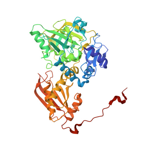
    7P9Q

  • PubMed Abstract: 

    The ubiquitous UbiD family of reversible decarboxylases is implicated in a wide range of microbial processes and depends on the prenylated flavin mononucleotide cofactor for catalysis. However, only a handful of UbiD family members have been characterized in detail, and comparison between these has suggested considerable variability in enzyme dynamics and mechanism linked to substrate specificity. In this study, we provide structural and biochemical insights into the indole-3-carboxylic acid decarboxylase, representing an UbiD enzyme activity distinct from those previously studied. Structural insights from crystal structure determination combined with small-angle X-ray scattering measurements reveal that the enzyme likely undergoes an open-closed transition as a consequence of domain motion, an event that is likely coupled to catalysis. We also demonstrate that the indole-3-carboxylic acid decarboxylase can be coupled with carboxylic acid reductase to produce indole-3-carboxyaldehyde from indole + CO 2 under ambient conditions. These insights provide further evidence for a common mode of action in the widespread UbiD enzyme family.


  • Organizational Affiliation
    • Manchester Institute of Biotechnology, University of Manchester, Manchester, UK.

Macromolecule Content 

  • Total Structure Weight: 306.23 kDa 
  • Atom Count: 21,468 
  • Modeled Residue Count: 2,714 
  • Deposited Residue Count: 2,766 
  • Unique protein chains: 1

Macromolecules

Find similar proteins by:|  3D Structure
Entity ID: 1
MoleculeChains  Sequence LengthOrganismDetailsImage
AnInD
A, B, C, D, E
A, B, C, D, E, F
461Glutamicibacter nicotianaeMutation(s): 0 

Small Molecules

Ligands 3 Unique
IDChains Name / Formula / InChI Key2D Diagram3D Interactions
4LU
(Subject of Investigation/LOI)

Query on 4LU


Download:Ideal Coordinates CCD File
I [auth A]
L [auth B]
O [auth C]
R [auth D]
U [auth E]
I [auth A],
L [auth B],
O [auth C],
R [auth D],
U [auth E],
X [auth F]
1-deoxy-5-O-phosphono-1-(3,3,4,5-tetramethyl-9,11-dioxo-2,3,8,9,10,11-hexahydro-7H-quinolino[1,8-fg]pteridin-12-ium-7-y l)-D-ribitol
C22 H30 N4 O9 P
KOUJZPGFPGLHCZ-IYOUNJFTSA-O
MN

Query on MN


Download:Ideal Coordinates CCD File
H [auth A]
K [auth B]
N [auth C]
Q [auth D]
T [auth E]
H [auth A],
K [auth B],
N [auth C],
Q [auth D],
T [auth E],
W [auth F]
MANGANESE (II) ION
Mn
WAEMQWOKJMHJLA-UHFFFAOYSA-N
NA

Query on NA


Download:Ideal Coordinates CCD File
G [auth A]
J [auth B]
M [auth C]
P [auth D]
S [auth E]
G [auth A],
J [auth B],
M [auth C],
P [auth D],
S [auth E],
V [auth F]
SODIUM ION
Na
FKNQFGJONOIPTF-UHFFFAOYSA-N

Experimental Data & Validation

Experimental Data

  • Method: X-RAY DIFFRACTION
  • Resolution: 2.53 Å
  • R-Value Free:  0.221 (Depositor), 0.224 (DCC) 
  • R-Value Work:  0.184 (Depositor), 0.189 (DCC) 
  • R-Value Observed: 0.186 (Depositor) 
Space Group: P 1 21 1
Unit Cell:
Length ( Å )Angle ( ˚ )
a = 104.2α = 90
b = 195.45β = 116.35
c = 105.21γ = 90
Software Package:
Software NamePurpose
REFMACrefinement
XDSdata reduction
Aimlessdata scaling
PHASERphasing
PDB_EXTRACTdata extraction

Structure Validation

View Full Validation Report


Ligand Structure Quality Assessment 


Entry History 

& Funding Information

Deposition Data


Funding OrganizationLocationGrant Number
European Research Council (ERC)United Kingdom--

Revision History  (Full details and data files)

  • Version 1.0: 2022-03-02
    Type: Initial release
  • Version 1.1: 2022-09-14
    Changes: Database references
  • Version 1.2: 2024-01-31
    Changes: Data collection, Refinement description